. 2023 Dec;10(12):2238-2254.
doi: 10.1002/acn3.51908. Epub 2023 Sep 30.

Dynamic causal modeling of reorganization of memory and language networks in temporal lobe epilepsy

Affiliations

Dynamic causal modeling of reorganization of memory and language networks in temporal lobe epilepsy

Alireza Fallahi et al. Ann Clin Transl Neurol. 2023 Dec.

Abstract

Objective: To evaluate the alterations of language and memory functions using dynamic causal modeling, in order to identify the epileptogenic hemisphere in temporal lobe epilepsy (TLE).

Methods: Twenty-two patients with left TLE and 13 patients with right TLE underwent functional magnetic resonance imaging (fMRI) during four memory and four language mapping tasks. Dynamic causal modeling (DCM) was employed on fMRI data to examine effective directional connectivity in memory and language networks and the alterations in people with TLE compared to healthy individuals.

Results: DCM analysis suggested that TLE can influence the memory network more widely compared to the language network. For memory mapping, it demonstrated overall hyperconnectivity from the left hemisphere to the other cranial regions in the picture encoding, and from the right hemisphere to the other cranial regions in the word encoding tasks. On the contrary, overall hypoconnectivity was seen from the brain hemisphere contralateral to the seizure onset in the retrieval tasks. DCM analysis further manifested hypoconnectivity between the brain's hemispheres in the language network in patients with TLE compared to controls. The CANTAB® neuropsychological test revealed a negative correlation for the left TLE and a positive correlation for the right TLE cohorts for the connections extracted by DCM that were significantly different between the left and right TLE cohorts.

Interpretation: In this study, dynamic causal modeling evidenced the reorganization of language and memory networks in TLE that can be used for a better understanding of the effects of TLE on the brain's cognitive functions.


Conflict of interest statement

There is no conflict of interest for any of the authors to disclose.

Figures

Figure 1
Figure 1
Sample task design for memory and language mapping.
Figure 2
Figure 2
Memory and language network nodes. FP, Frontal pole; L‐ANG, left angular; L‐EC, left EC; L‐HIP, left hippocampus; L‐IFG_PO, left inferior frontal gyrus, pars opercularis; L‐IFG_PT, left inferior frontal gyrus, pars triangularis; L‐MFG, left middle frontal gyrus; L‐MTG, left middle temporal gyrus; L‐PHIP, left Para Hippocampus; L‐PMTG, left posterior middle temporal gyrus; L‐PRC, left PRC; L‐PSG, left posterior supramarginal gyrus; L‐RSC, left RSC; L‐THA, left thalamus; L‐TP, left temporal pole; MPFC, middle PFC; R‐ANG, right angular; R‐EC, right EC; R‐HIP, right hippocampus; R‐PHIP, right para hippocampus; R‐PRC, right PRC; R‐PSTG, right posterior superior temporal gyrus; R‐RSC, right RSC; R‐THA, right thalamus; R‐TP, right temporal pole; SFG_1, superior frontal gyrus; SFG_2, superior frontal gyrus.
Figure 3
Figure 3
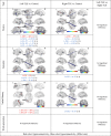
Result of significant difference in effective connectivity between left‐TLE, right‐TLE, and control groups using memory mapping tasks in picture encoding and retrieval tasks. L‐ANG, left angular; L‐EC, left EC; L‐HIP, left hippocampus; L‐PHIP, left para hippocampus; L‐PRC, left PRC; L‐RSC, left RSC; L‐THA, left thalamus; MPFC, middle PFC; R‐ANG, RIGHT ANGULAR; R‐EC, right EC; R‐HIP, right hippocampus; R‐PHIP, right para hippocampus; R‐PRC, right PRC; R‐RSC, right RSC; R‐THA, right Thalamus.
Figure 4
Figure 4
Result of significant difference in effective connectivity between left‐TLE, right‐TLE, and control groups using memory mapping tasks in word encoding and retrieval tasks. L‐ANG, left angular; L‐EC, left EC; L‐HIP, left hippocampus; L‐PHIP, left para hippocampus; L‐PRC, left PRC; L‐RSC, left RSC; L‐THA, left thalamus; MPFC, middle PFC; R‐ANG, right angular; R‐EC, right EC; R‐HIP, right hippocampus; R‐PHIP, right para hippocampus; R‐PRC, right PRC; R‐RSC, right RSC; R‐THA, right thalamus.
Figure 5
Figure 5
Bar graph for significant connection between Left‐TLE and Right‐TLE groups for memory mapping tasks. Red graphs represent the Left‐TLE group, yellow graphs represent the Right‐TLE group and blue graphs represent the HC group.
Figure 6
Figure 6
Result of significant difference in effective connectivity between left‐TLE, right‐TLE, and control groups using language mapping task. FP, frontal pole; L‐IFG_PO, left inferior frontal gyrus, pars opercularis; L‐IFG_PT, left inferior frontal gyrus, pars triangularis; L‐MFG, left middle frontal gyrus; L‐MTG, left middle temporal gyrus; L‐PMTG, left posterior middle temporal gyrus; L‐PSG, left posterior supramarginal gyrus; L‐TP, left temporal pole; R‐PSTG, right posterior superior temporal gyrus; R‐TP, right temporal pole; SFG_1, superior frontal gyrus; SFG_2, superior frontal gyrus.
Figure 7
Figure 7
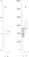
Box plot representation of CANTAB PAL and PRM tests with a significant difference between left and right TLE groups. The red stars denote significant differences between the two groups (t‐test P‐value < 0.05). L, left TLE group; PAL SCFT, PAL stages completed on the first trial; PRM MCL, PRM mean correct latency; R, right TLE group.
Figure 8
Figure 8
Correlation between significant connections between the left and right TLE groups and CANTAB PAL and PRM tests in picture encoding and retrieval tasks. Only correlations that have significant linear relations are represented. Blue colors and green colors are related to the left and right TLE groups, respectively. LHIP, left hippocampus; LPHIP, left parahippocampus; REC, right entorhinal cortex; RHIP, right hippocampus.
Figure 9
Figure 9
Correlation between significant connections between the left and right TLE groups and CANTAB PAL and PRM tests in word encoding and retrieval tasks. Only correlations that have significant linear relations are represented. Blue colors and green colors are related to the left and right TLE groups, respectively. LANG, Left angular; LPRC, left perirhinal cortex; LRSC, left retrosplenial cortex; LTHA, left thalamus; REC, right entorhinal cortex; RRSC, right retrosplenial cortex; RPRC, right perirhinal cortex.

References

    1. Wiebe S, Blume WT, Girvin JP, Eliasziw M. A randomized, controlled trial of surgery for temporal‐lobe epilepsy. N Engl J Med. 2001;345:311‐318. - PubMed
    1. Berg AT, Scheffer IE. New concepts in classification of the epilepsies: entering the 21st century. Epilepsia. 2011;52:1058‐1062. - PubMed
    1. Alessio A, Pereira FRS, Sercheli MS, et al. Brain plasticity for verbal and visual memories in patients with mesial temporal lobe epilepsy and hippocampal sclerosis: an fMRI study. Hum Brain Mapp. 2013;34:186‐199. - PMC - PubMed
    1. Jaimes‐Bautista AG, Rodríguez‐Camacho M, Martínez‐Juárez IE, Rodríguez‐Agudelo Y. Semantic processing impairment in patients with temporal lobe epilepsy. Epilepsy Res Treat. 2015;2015:1‐8. - PMC - PubMed
    1. Metternich B, Buschmann F, Wagner K, Schulze‐Bonhage A, Kriston L. Verbal fluency in focal epilepsy: a systematic review and meta‐analysis. Neuropsychol Rev. 2014;24:200‐218. - PubMed

Publication types

LinkOut - more resources