. 2023 Oct 11;31(10):1639-1654.e10.
doi: 10.1016/j.chom.2023.08.018. Epub 2023 Sep 29.

Iron acquisition by a commensal bacterium modifies host nutritional immunity during Salmonella infection

Affiliations

Iron acquisition by a commensal bacterium modifies host nutritional immunity during Salmonella infection

Luisella Spiga et al. Cell Host Microbe. .

Abstract

During intestinal inflammation, host nutritional immunity starves microbes of essential micronutrients, such as iron. Pathogens scavenge iron using siderophores, including enterobactin; however, this strategy is counteracted by host protein lipocalin-2, which sequesters iron-laden enterobactin. Although this iron competition occurs in the presence of gut bacteria, the roles of commensals in nutritional immunity involving iron remain unexplored. Here, we report that the gut commensal Bacteroides thetaiotaomicron acquires iron and sustains its resilience in the inflamed gut by utilizing siderophores produced by other bacteria, including Salmonella, via a secreted siderophore-binding lipoprotein XusB. Notably, XusB-bound enterobactin is less accessible to host sequestration by lipocalin-2 but can be "re-acquired" by Salmonella, allowing the pathogen to evade nutritional immunity. Because the host and pathogen have been the focus of studies of nutritional immunity, this work adds commensal iron metabolism as a previously unrecognized mechanism modulating the host-pathogen interactions and nutritional immunity.

Keywords: Salmonella; commensal iron metabolism; enteric pathogen; gut microbiota resilience; intestinal inflammation; nutritional immunity; siderophore.


Conflict of interest statement

Declaration of interests The authors declare no competing interests.

Figures

Figure 1:
Figure 1:. XusB is a surface-exposed protein that is enriched in outer membrane vesicles
(A) Schematic representation of the xusABC operon in B. thetaiotaomicron VPI-5482. (B) Sequence features of XusB N-terminus. (C) B. thetaiotaomicron cells were cultured in iron-limiting media and treated with indicated concentrations of protease K. Degradation of HA-tagged XusB on whole B. thetaiotaomicron cells was assessed via western blots. (D) B. thetaiotaomicron cells were cultured in iron-limiting media and fractionated into outer membrane vesicles (OMV-P), cell-free supernatant (OMV-S), membrane (M), and cytoplasm/periplasm (C) fractions. These fractions were then mixed with identical fractions of B. thetaiotaomicron strains expressing HA-tagged OmpF or Flag-tagged BF1581 and resolved on SDS-PAGE gel. RpoB, OmpF, and BF1581 are cytoplasmic, membrane, and OMV controls, respectively.
Figure 2:
Figure 2:. Bacteroidetes encode homologs of XusB, which binds to enterobactin with high affinity
(A) Size exclusion chromatography traces for recombinant XusB (280 nm) incubated with Fe-enterobactin (550 nm). (B) Changes of intrinsic tryptophan fluorescence (Δ fluorescence) in XusB upon binding to enterobactin. The dissociation constant was derived by fitting Ent concentrations vs. fluorescence intensity changes to a one-site specific binding model. (C) The overall structure of XusBΔN. N- and C-termini are indicated, and blades are numbered. (D) Overall electrostatic profile of XusB docked with Fe-Ent. Enterobactin is shown as sticks with carbon atoms colored in green, nitrogen in blue, and oxygen in red, with the Fe3+ represented as an orange sphere. The color scale represents electrostatic potentials in units of kT/e ranging from −5 (red, negatively charged) to +5 (blue, positively charged). (E) Close-up of XusBΔN residues interacting with Fe3+-Ent. Residues and catechol arms of Fe3+-Ent are numbered. (F) B. thetaiotaomicron strains were cultured in BHIS medium, followed by subculturing in iron-limited (200 μmol/L of BPS) SDM supplemented with 0.5 μM enterobactin. Growth of B. thetaiotaomicron was determined by measuring OD600. (G) Overlay of ΔN-terminus structures of XusB and its homologs. Bars represent the geometric mean. ***, P < 0.001.
Figure 3:
Figure 3:. XusB can function as an early step in xenosiderophore acquisition
(A-C) Supernatant from donor B. thetaiotaomicron strains grown in iron-limited medium (BPS-supplemented SDM) was filter-sterilized, loaded with Fe-Ent, ultracentrifuged to collect OMV fraction, washed to remove unbound ligand, and introduced to recipient cells in BPS-supplemented SDM. (A) Schematic representation and (B-C) Bacterial growth measured by OD600. (D) The B. thetaiotaomicron wild-type strain and an isogenic ΔxusB mutant were cultured in iron-deprived media to exhaust endogenous iron before subcultured in the presence of Fe-Ent loaded OMVs collected from indicated strains. ICP-MS was used to assess cellular iron levels. Bars represent the geometric mean. **, P < 0.01; ***, P < 0.001; n.s., not statistically significant.
Figure 4:
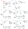
Figure 4:. Role of xusB in B. thetaiotaomicron resilience during murine S. Tm infection
(A-E) Groups of C57BL/6 mice were treated with a cocktail of antibiotics, followed by intragastrical inoculation of (A-B) the B. thetaiotaomicron wild-type strain, or (C-E) an equal mixture of the B. thetaiotaomicron wild-type strain and an isogenic mutant (ΔxusB), or a strain expressing the enterobactin-binding deficient mutant (ΔxusB xusBentMT). Mice were then either mock-treated, intragastrically challenged with S. Tm SL1344, or an isogenic entB mutant (N = 5-6/group). (A&C) Schematic presentation of the experiments. Four days after infection, the cecal contents were collected, (B) the iron concentrations of indicated fractions were quantified using ICP-MS, and the abundance of (D) B. thetaiotaomicron and (E) S. Tm was determined by plating on selective agar. The competitive index was calculated as the ratio of the two strains in the cecal content, corrected by the ratio in the inoculum. (F-H) Groups of C57BL/6 mice were treated with a cocktail of antibiotics, followed by intragastrical inoculation of either the B. thetaiotaomicron wild-type strain or an isogenic ΔxusB mutant. Mice were challenged with indicated S. Tm SL1344 strains for 4 days (N = 10-12/group). The abundance of each B. thetaiotaomicron (G) and S. Tm (H) strain in cecal contents was determined by plating on selective agar. Bars represent the geometric mean. *, P < 0.05; **, P < 0.01; ***; P < 0.001; n.s., not statistically significant.
Figure 5:
Figure 5:. Role of the XusB in interspecies competition for siderophores
(A) Model schematic of XusB in modifying enterobactin accessibility. (B) B. thetaiotaomicron or B. vulgatus were inoculated into iron-starved, semi-defined medium (200 μmol/L BPS) supplemented with either free Fe-Ent or Fe-Ent complex with recombinant XusB; bacterial growth was measured 48 hours post-inoculation by OD600. (C-F) Filter-sterilized supernatant from donor B. thetaiotaomicron strains grown in iron-limited medium (C&E) or indicated recombinant XusB protein (D&F) was loaded with Fe-Ent, washed to remove unbound ligand, and introduced to recipient S. Tm (C-D) or E. coli (E-F) strains grown in SDM supplemented with 200 μmol/L BPS. Bacteria were cultured for 48 hours, and bacterial growth was measured by OD600. Bars represent the geometric mean. **; P < 0.01; ***; P < 0.001; ns, not statistically significant.
Figure 6:
Figure 6:. XusB modulates the interactions between S. Tm and lipocalin-2 in vitro
(A-C) An equal mixture of S. Tm wild-type strain and an isogenic ΔfepA iroN mutant was inoculated into SDM supplemented with BPS and recombinant lipolicalin-2, in the absence or presence of XusB, for 16 hours. Competition index for the S. Tm SL1344 wild-type strain vs. the ΔfepA iroN mutant strain in the presence of (B) recombinant XusB and (C) XusB-containing OMVs was calculated as the ratio of the two strains in the culture, corrected by the ratio in the inoculum. Chelex: Pretreatment of divalent ion chelating agent Chelex 100. HI: Heat inactivation. Bars represent the geometric mean. **, P < 0.01; ***, P < 0.001; n.s., not statistically significant.
Figure 7:
Figure 7:. Role of XusB in pathogen-host nutritional immunity interactions during S. Tm infection
(A-B) Groups of gnotobiotic Swiss Webster mice were monoassociated with either the B. thetaiotaomicron wild-type strain or the isogenic ΔxusB mutant for 7 days. Mice were then challenged with the S. Tm SL1344 for 2 days. The outer membrane vesicle fraction of the intestinal contents was collected, and the proteome was profiled. (A) Schematic representation. (B) XusB peptide abundance measured by UHPLC-MS/MS. (C-D) Groups of C57BL/6 mice were treated with a cocktail of antibiotics, followed by intragastrical inoculation of indicated B. thetaiotaomicron strains (N = 10-12/group). Mice were inoculated with an equal mixture of the S. Tm SL1344 wild-type strain and the isogenic ΔfepA iroN mutant strain. (C) Schematic representation. 4 days later, cecal contents were collected, and the competitive index (D) for the S. Tm SL1344 wild-type strain vs. the ΔfepA iroN mutant strain was determined. (E-F) Groups of antibiotic-pretreated C57BL/6 mice were intragastrically inoculated with a ΔxusB mutant strain. Two days later, mice were inoculated with an equal mixture of the S. Tm SL1344 wild-type strain and the isogenic ΔfepA iroN mutant strain, followed by intragastrical administration of WT-OMVs derived ΔxusB-OMV(N = 10-11/group) daily. (E) Schematic representation. 4 days later, cecal contents were collected, and the competition index (F) for the S. Tm SL1344 wild-type strain vs. the ΔfepA iroN mutant strain was determined. (G-I) Groups of gnotobiotic Swiss Webster mice were monoassociated with either the B. thetaiotaomicron wild-type strain or the ΔxusB mutant (N = 11-12/group) for 7 days. Mice were then challenged with an equal mixture of the S. Tm SL1344 wild-type strain and the ΔfepA iroN mutant strain for 3 days. (G) Schematic representation of the experiment. The abundance of B. thetaiotaomicron (H) and the competition index (I) for S. Tm SL1344 wild-type strain vs. the ΔfepA iroN mutant strain in the cecal contents were determined. (J-M) Groups of C57BL/6 mice were treated with a single oral dose of streptomycin, followed by intragastrical inoculation of (J-K) the S. Tm SL1344 ΔiroB mutant or (L-M) the S. Tm wild-type strain. Mice also received daily administration of equal amounts of WT-OMVs or ΔxusB-OMV (N = 5-11/group). (J&L) Schematic representation of the experiment. 2 days later, cecal contents were collected, and the population (K&M) for the S. Tm strains was determined. Bars represent the geometric mean. *, P < 0.05; **, P < 0.01; n.s., not statically significant.

Update of

Comment in

  • Robbing the thief.
    Jaswal K, Behnsen J. Jaswal K, et al. Cell Host Microbe. 2023 Oct 11;31(10):1597-1599. doi: 10.1016/j.chom.2023.09.009. Cell Host Microbe. 2023. PMID: 37827119

References

    1. Almeida A, Mitchell AL, Boland M, Forster SC, Gloor GB, Tarkowska A, Lawley TD, and Finn RD (2019). A new genomic blueprint of the human gut microbiota. Nature 568, 499–504. 10.1038/s41586-019-0965-1. - DOI - PMC - PubMed
    1. Vemuri R, Shankar EM, Chieppa M, Eri R, and Kavanagh K (2020). Beyond Just Bacteria: Functional Biomes in the Gut Ecosystem Including Virome, Mycobiome, Archaeome and Helminths. Microorganisms 8. 10.3390/microorganisms8040483. - DOI - PMC - PubMed
    1. Sommer F, Anderson JM, Bharti R, Raes J, and Rosenstiel P (2017). The resilience of the intestinal microbiota influences health and disease. Nat Rev Microbiol 15, 630–638. 10.1038/nrmicro.2017.58. - DOI - PubMed
    1. Scheffer M, Carpenter S, Foley JA, Folke C, and Walker B (2001). Catastrophic shifts in ecosystems. Nature 413, 591–596. 10.1038/35098000. - DOI - PubMed
    1. Ingrisch J, and Bahn M (2018). Towards a Comparable Quantification of Resilience. Trends Ecol Evol 33, 251–259. 10.1016/j.tree.2018.01.013. - DOI - PubMed

Key Resource Table Reference:

    1. Koropatkin NM, et al., Starch catabolism by a prominent human gut symbiont is directed by the recognition of amylose helices. Structure, 2008. 16(7): p. 1105–15. - PMC - PubMed
    1. Zhu W, et al., Xenosiderophore Utilization Promotes Bacteroides thetaiotaomicron Resilience during Colitis. Cell Host Microbe, 2020. 27(3): p. 376–388 e8. - PMC - PubMed
    1. Song YL, et al., “Bacteroides nordii” sp. nov. and “Bacteroides salyersae” sp. nov. isolated from clinical specimens of human intestinal origin. J Clin Microbiol, 2004. 42(12): p. 5565–70. - PMC - PubMed
    1. Hoiseth SK and Stocker BA, Aromatic-dependent Salmonella typhimurium are non-virulent and effective as live vaccines. Nature, 1981. 291(5812): p. 238–9. - PubMed
    1. Pal D, et al., Multipartite regulation of rctB, the replication initiator gene of Vibrio cholerae chromosome II. J Bacteriol, 2005. 187(21): p. 7167–75. - PMC - PubMed

LinkOut - more resources