Review
. 2023 Sep 30;28(1):76.
doi: 10.1186/s11658-023-00489-y.

Oxidative stress as a key modulator of cell fate decision in osteoarthritis and osteoporosis: a narrative review

Affiliations
Review

Oxidative stress as a key modulator of cell fate decision in osteoarthritis and osteoporosis: a narrative review

Jana Riegger et al. Cell Mol Biol Lett. .

Abstract

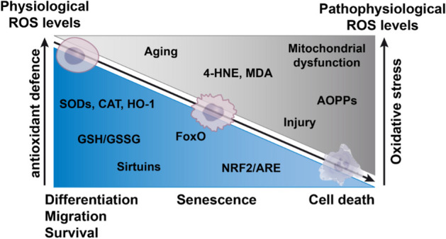
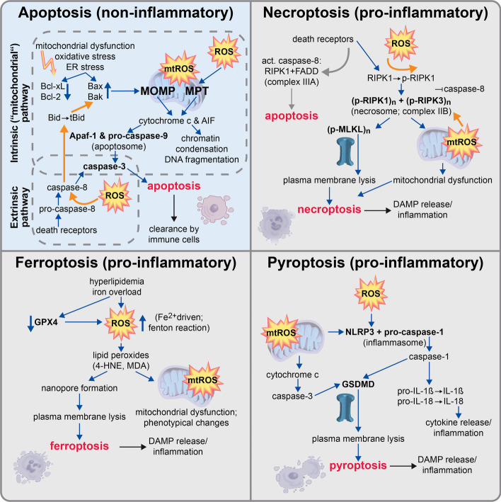
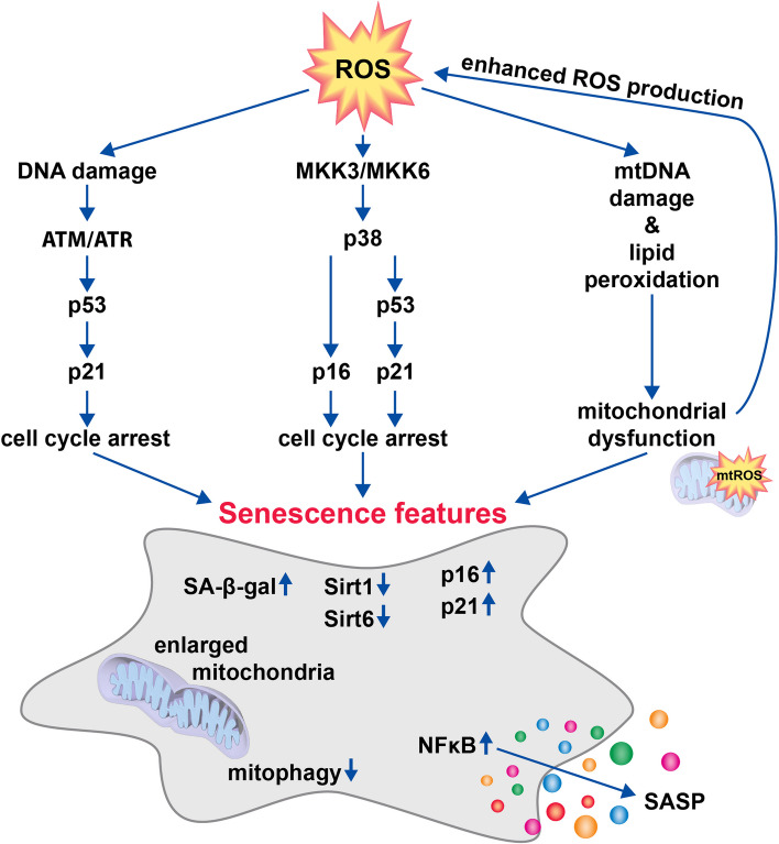
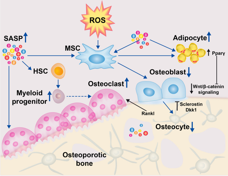
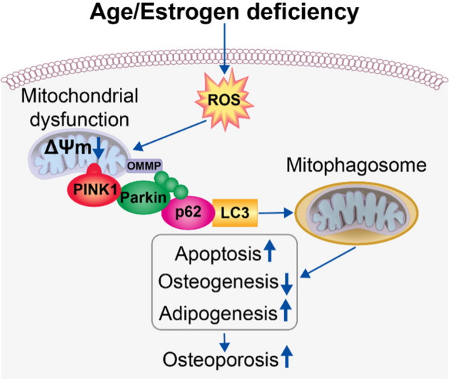
During aging and after traumatic injuries, cartilage and bone cells are exposed to various pathophysiologic mediators, including reactive oxygen species (ROS), damage-associated molecular patterns, and proinflammatory cytokines. This detrimental environment triggers cellular stress and subsequent dysfunction, which not only contributes to the development of associated diseases, that is, osteoporosis and osteoarthritis, but also impairs regenerative processes. To counter ROS-mediated stress and reduce the overall tissue damage, cells possess diverse defense mechanisms. However, cellular antioxidative capacities are limited and thus ROS accumulation can lead to aberrant cell fate decisions, which have adverse effects on cartilage and bone homeostasis. In this narrative review, we address oxidative stress as a major driver of pathophysiologic processes in cartilage and bone, including senescence, misdirected differentiation, cell death, mitochondrial dysfunction, and impaired mitophagy by illustrating the consequences on tissue homeostasis and regeneration. Moreover, we elaborate cellular defense mechanisms, with a particular focus on oxidative stress response and mitophagy, and briefly discuss respective therapeutic strategies to improve cell and tissue protection.

Keywords: Bone; Cartilage; Cell death; Cell fate decision; Mitochondrial dysfunction; Osteoarthritis; Osteoporosis; Oxidative stress; ROS; Senescence.


Conflict of interest statement

The authors declare no competing interests.

Figures

Fig. 1
Fig. 1
Overview of the redox balance in cell fate decision. The decrease of cellular antioxidant defense mechanisms (blue area) results in an increase of oxidative stress, driven and characterized by various factors (gray area). Alterations in the redox balance differentially effect cellular behavior and fate, ranging from physiological to pathophysiological processes
Fig. 2
Fig. 2
ROS involvement in regulated cell death. While apoptotic cell death (blue background) is considered as a largely silent and non-inflammatory form of cell death, other forms such as necroptosis, ferroptosis, and pyroptosis (gray background) are thought to promote a pro-inflammatory response. Regardless of the pathophysiologic consequences, ROS and mitochondrial dysfunction (mtROS) play a decisive role in the execution of all described forms of regulated cell death. Apaf-1 apoptosis activating factor-1, DAMP damage-associated molecular patterns, GPX4 glutathione peroxidase 4, GSDMD gasdermins, MOMP mitochondrial outer membrane permeabilization, MPT mitochondrial permeability transition, (mt)ROS mitochondrial) reactive oxygen species, NLRP3 nucleotide-binding domain and leucine-rich repeat protein-3
Fig. 3
Fig. 3
ROS as a driver of senescence. Enhanced ROS levels result in cellular damage and consequent cell cycle arrest mediated via p53, p21, and p16. Moreover, ROS damages mitochondrial DNA (mtDNA) and causes lipid peroxidation initiating mitochondrial dysfunction and thus lead to enhanced ROS generation. Other senescence features are the expression of senescence-associated β-galactosidase (SA-β-Gal), downregulation of Sirt1 and Sirt6, upregulation of p21 and p16, presence of enlarged mitochondria, inhibition of mitophagy, and secretion of senescence-associated secretory phenotype (SASP) factors mainly driven by the NFκB
Fig. 4
Fig. 4
Role of ROS in bone cell fate regulation. ROS (reactive oxygen species) generation due to aging and estrogen deficiency is accompanied by increased cellular senescence characterized by the senescence-associated secretory phenotype (SASP) in the bone tissue. The SASP facilitates the accumulation of adipocytes in the bone marrow at the expense of osteoblast formation by inducing peroxisome proliferator activated receptor gamma (Pparγ) in mesenchymal progenitor cells (MSCs). Additionally, the SASP promotes myeloid progenitor development from the hematopoietic stem cell (HSC) lineage. Consequently, an elevated monocyte level is associated with an increase of osteoclast formation. Apoptotic osteocytes are involved in increased bone resorption by producing an unbalanced RANKL level. Moreover, apoptotic osteocytes induce sclerostin and Dickkopf-related protein (Dkk) 1 production, which inhibits Wnt/b-catenin mediated osteogenesis
Fig. 5
Fig. 5
Role of ROS in regulating mitophagy in OP. Age and estrogen deficiency cause enhanced reactive oxygen species (ROS) generation in MSCs. ROS lead to mitochondrial dysfunction accompanied by a reduced mitochondrial membrane potential (ΔΨm). Subsequent PINK1 accumulation at the outer surface of the mitochondrial membrane initiates mitophagy by recruitment of Parkin that promotes ubiquitination (green dots) of OMM proteins (OMMP). Ubiquitinated proteins bind directly or indirectly to autophagy receptor LC3 via p62 on mitophagosomes. Aberrant mitophagy may result in increased apoptosis, promoting marrow adipogenesis as well as suppressing osteogenesis, all consequently contributing to OP development

Similar articles

Cited by

References

    1. Balaban RS, Nemoto S, Finkel T. Mitochondria, oxidants, and aging. Cell. 2005;120(4):483–495. doi: 10.1016/j.cell.2005.02.001. - DOI - PubMed
    1. Zullo A, Guida R, Sciarrillo R, Mancini FP. Redox homeostasis in cardiovascular disease: the role of mitochondrial sirtuins. Front Endocrinol (Lausanne). 2022;13:858330. doi: 10.3389/fendo.2022.858330. - DOI - PMC - PubMed
    1. Nathan C, Cunningham-Bussel A. Beyond oxidative stress: an immunologist’s guide to reactive oxygen species. Nat Rev Immunol. 2013;13(5):349–361. doi: 10.1038/nri3423. - DOI - PMC - PubMed
    1. Schieber M, Chandel NS. ROS function in redox signaling and oxidative stress. Curr Biol. 2014;24(10):R453–R462. doi: 10.1016/j.cub.2014.03.034. - DOI - PMC - PubMed
    1. Sha Y, Marshall HE. S-nitrosylation in the regulation of gene transcription. Biochim Biophys Acta. 2012;1820(6):701–711. doi: 10.1016/j.bbagen.2011.05.008. - DOI - PMC - PubMed

Substances