. 2023 Sep 30;20(1):221.
doi: 10.1186/s12974-023-02907-6.

Receptor-interacting protein kinase 2 (RIPK2) profoundly contributes to post-stroke neuroinflammation and behavioral deficits with microglia as unique perpetrators

Affiliations

Receptor-interacting protein kinase 2 (RIPK2) profoundly contributes to post-stroke neuroinflammation and behavioral deficits with microglia as unique perpetrators

Jonathan Larochelle et al. J Neuroinflammation. .

Abstract

Background: Receptor-interacting protein kinase 2 (RIPK2) is a serine/threonine kinase whose activity propagates inflammatory signaling through its association with pattern recognition receptors (PRRs) and subsequent TAK1, NF-κB, and MAPK pathway activation. After stroke, dead and dying cells release a host of damage-associated molecular patterns (DAMPs) that activate PRRs and initiate a robust inflammatory response. We hypothesize that RIPK2 plays a damaging role in the progression of stroke injury by enhancing the neuroinflammatory response to stroke and that global genetic deletion or microglia-specific conditional deletion of Ripk2 will be protective following ischemic stroke.

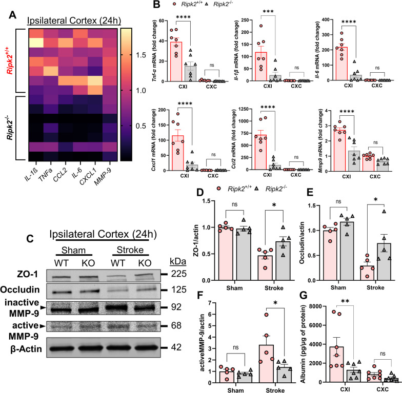
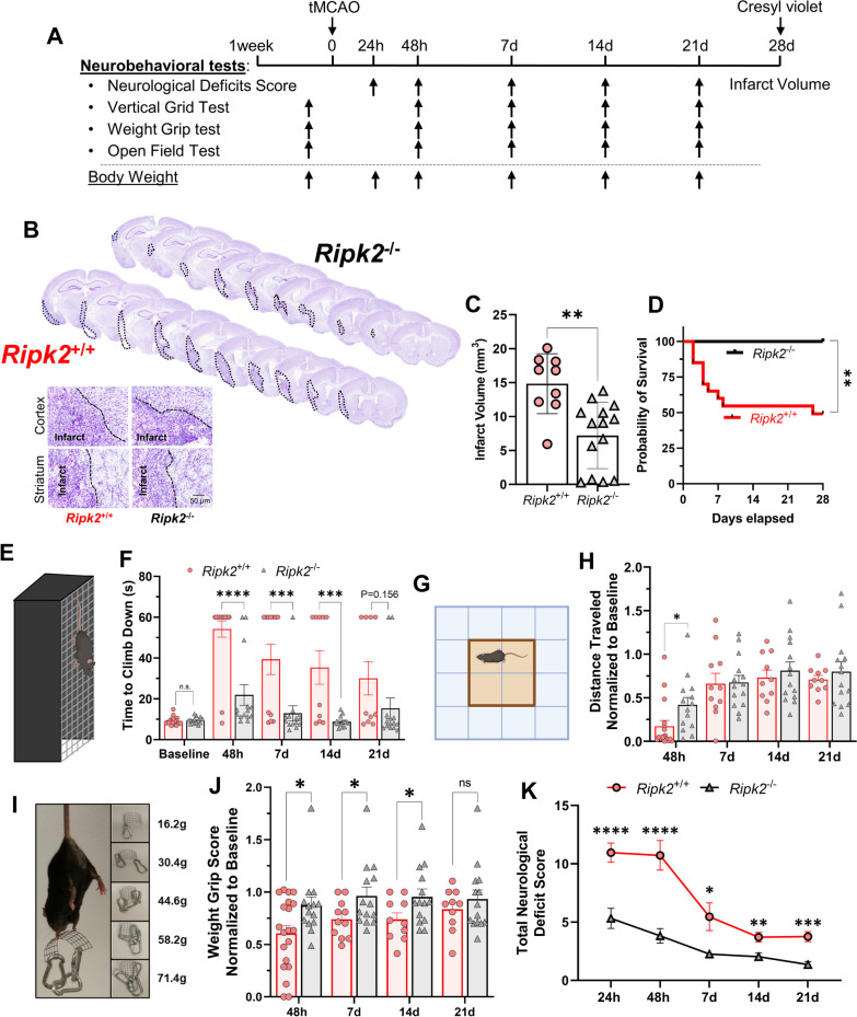
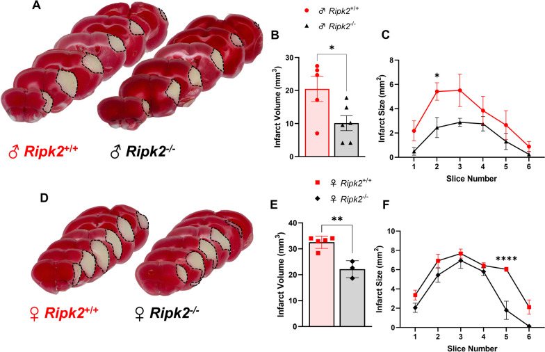
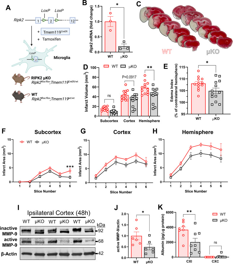
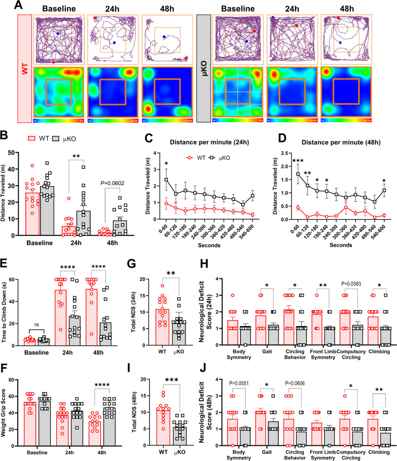
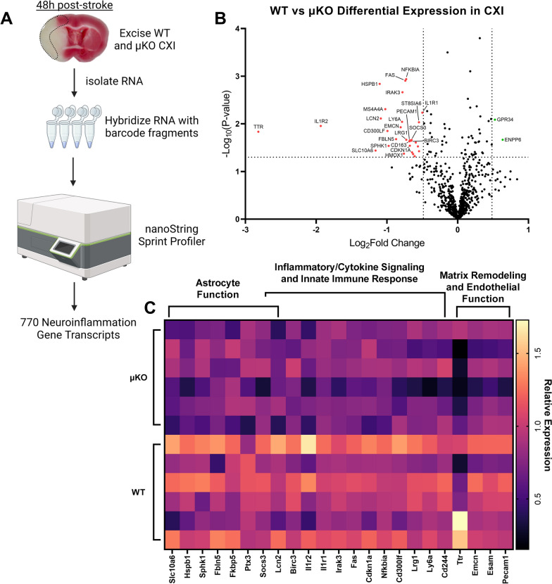
Methods: Adult (3-6 months) male mice were subjected to 45 min of transient middle cerebral artery occlusion (tMCAO) followed by 24 h, 48 h, or 28 days of reperfusion. Aged male and female mice (18-24 months) were subjected to permanent ischemic stroke and sacrificed 48 h later. Infarct volumes were calculated using TTC staining (24-48 h) or Cresyl violet staining (28d). Sensorimotor tests (weight grip, vertical grid, and open field) were performed at indicated timepoints. Blood-brain barrier (BBB) damage, tight junction proteins, matrix metalloproteinase-9 (MMP-9), and neuroinflammatory markers were assessed via immunoblotting, ELISA, immunohistochemistry, and RT-qPCR. Differential gene expression profiles were generated through bulk RNA sequencing and nanoString®.

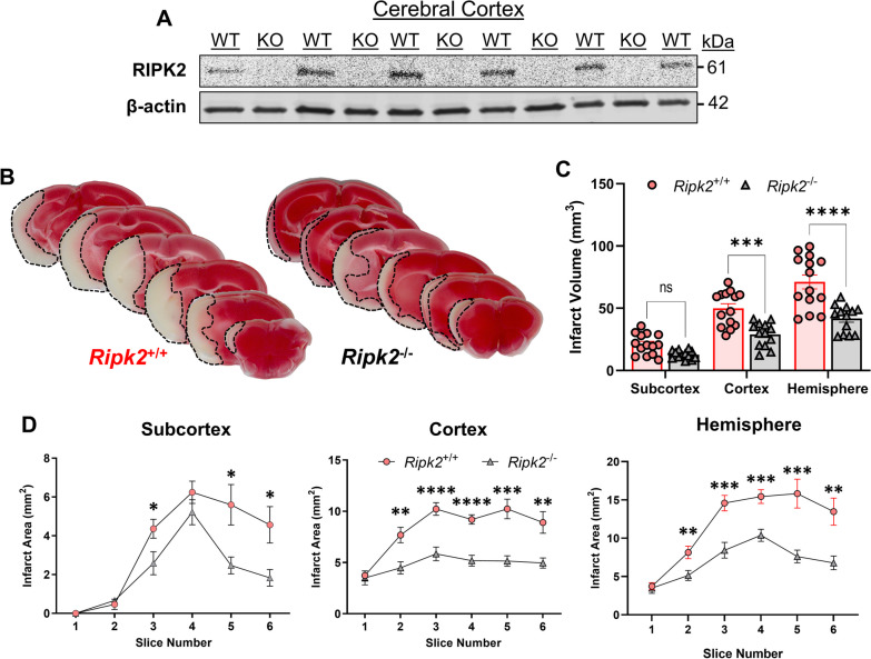
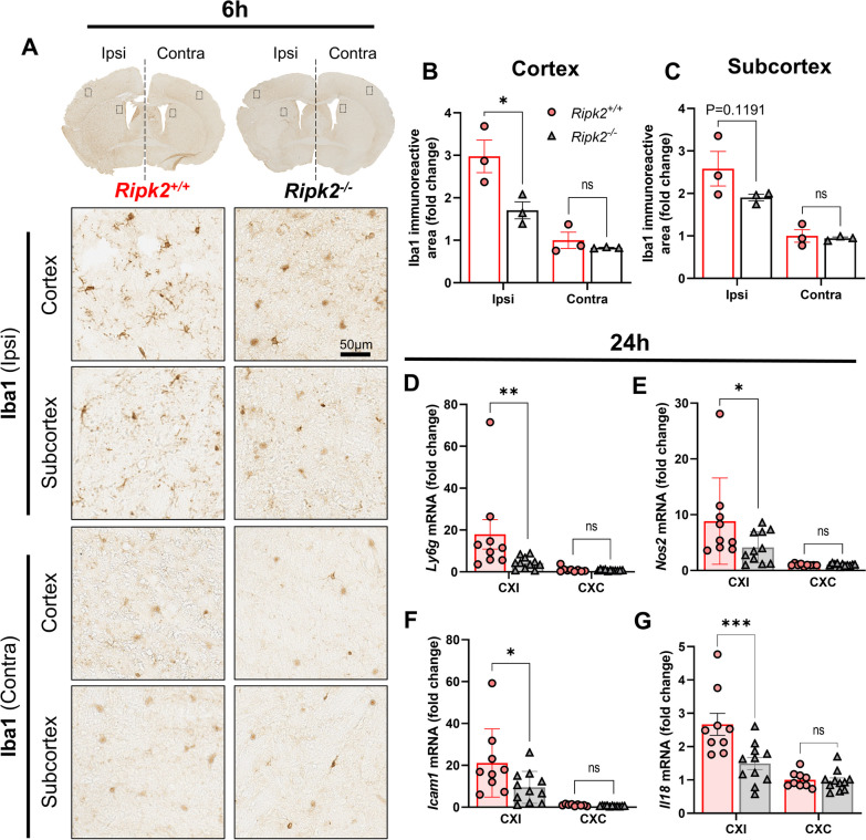
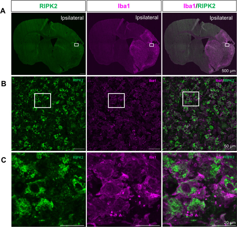
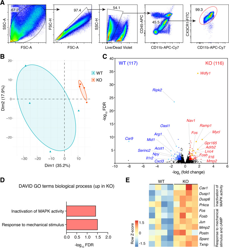
Results: Global genetic deletion of Ripk2 resulted in decreased infarct sizes and reduced neuroinflammatory markers 24 h after stroke compared to wild-type controls. Ripk2 global deletion also improved both acute and long-term behavioral outcomes with powerful effects on reducing infarct volume and mortality at 28d post-stroke. Conditional deletion of microglial Ripk2 (mKO) partially recapitulated our results in global Ripk2 deficient mice, showing reductive effects on infarct volume and improved behavioral outcomes within 48 h of injury. Finally, bulk transcriptomic profiling and nanoString data demonstrated that Ripk2 deficiency in microglia decreases genes associated with MAPK and NF-κB signaling, dampening the neuroinflammatory response after stroke injury by reducing immune cell activation and peripheral immune cell invasion.

Conclusions: These results reveal a hitherto unknown role for RIPK2 in the pathogenesis of ischemic stroke injury, with microglia playing a distinct role. This study identifies RIPK2 as a potent propagator of neuroinflammatory signaling, highlighting its potential as a therapeutic target for post-stroke intervention.

Keywords: Blood–brain barrier injury; Ischemic stroke; Microglia; Neuroinflammation; RIPK2.


Conflict of interest statement

The authors declare no competing interests.

Figures

Fig. 1
Fig. 1
Global deletion of Ripk2 dramatically reduces infarct volume after transient ischemic stroke. A Western blot (WB) of RIPK2 in the cerebral cortex homogenate from Ripk2+/+ (WT) and Ripk2−/− (KO) mice under naïve conditions. B Representative TTC staining of 1 mm-thick coronal brain slices from Ripk2+/+ and Ripk2−/− mice 24 h post-tMCAO. C Quantitative analysis of total infarction in each brain region. D Quantification of the infarction measured from each individual 1-mm-thick slice at the levels of the subcortex, cortex, and total hemisphere. A n = 5–6/group. BD Ripk2+/+: n = 14, Ripk2−/−: n = 13. Statistical differences determined by Student’s t test. *< 0.05, **< 0.01, ***P < 0.001, ****P < 0.0001
Fig. 2
Fig. 2
Ripk2−/− mice have dramatically lower pro-inflammatory activity and greater BBB preservation in the cerebral cortex 24 h post-stroke compared to Ripk2+/+ mice. A Heat map of inflammation-associated genes in the ipsilateral cerebral cortex comparing the difference in expression between the two genotypes. Horizontal lines denote individual animals, while vertical lines denote genes of interest. B Quantification of the heat map in (A), depicting mRNA levels of Tnfα, Il1β, Il6, Cxcl1, Ccl2, and Mmp9 in the ipsilateral (CXI) and contralateral (CXC) cortices of the two genotypes 24 h after stroke. C Western blot (WB) of the ipsilateral cortex depicting levels of ZO-1, Occludin, and Active-MMP-9, and β-Actin in Ripk2+/+ (WT) and Ripk2−/− (KO) brain homogenate. D–F Quantification of WB shown in C. G ELISA for the detection of the blood serum protein Albumin in the CXI and CXC of WT and KO mice 24 h after stroke, as a measurement of BBB permeability. A, B n = 7/genotype, (CF) n = 5/genotype, (G) n = 7/genotype. Statistical differences determined by two-way ANOVA, Turkey’s multiple comparison test. *P < 0.05, ***P < 0.001, ****P < 0.0001
Fig. 3
Fig. 3
Reduced immune cell infiltration and activation in Ripk2 deficient mice compared to controls. A Iba-1 staining of coronal brain sections 6 h after stroke injury with representative images of the ipsilateral (ipsi) and contralateral (contra) areas of interest. B, C Densitometric analysis and quantification of the area of Iba-1 staining in the cortex (B) and subcortex (C). D RT-qPCR for Nos2, Icam1, and Ly6g in the ipsi and contra cortices 24 h after stroke. AC n = 3/group. D n = 9–11/group. Differences determined by two-way ANOVA, multiple comparisons, Turkey’s multiple comparison test. *P < 0.05, ***P < 0.001
Fig. 4
Fig. 4
Ripk2−/− mice have smaller infarcts, zero mortality, and better behavioral outcomes 28-day post-stroke compared to Ripk2.+/+ mice. A Experimental design for the 28-day, longitudinal study. B Cresyl violet staining of 30 μm-thick brain slices at 28 days after stroke with inset depicting delineated areas of the infarct. C Quantification of infarct sizes from Cresyl violet-stained sections from each genotype. D Kaplan–Meier survival curve out to 28-day post-stroke. E, F Time required for mice to descend the vertical grid (E) after stroke, quantified in F. G, Distance traveled in the open field chamber (G) after stroke, with each value being normalized to that animal’s baseline distance (H). I, J Weight grip scores determined after stroke. Mice grip weights of increasing mass (I) and scores were quantified (J). K Total neurological deficit score assigned at indicated timepoints after stroke. B, C n = 9–14/genotype. FK n = 14–21/genotype at 24 h and 48 h, n = 11–14/genotype at 7d, n = 10–14/genotype at 14d and 21d. C Student’s t test. D Gehan–Breslow–Wilcoxon test. F, H Two-way ANOVA. J, K Mann–Whitney test. *P < 0.05, **P < 0.01, ***P < 0.001, ****P < 0.0001
Fig. 5
Fig. 5
Ripk2 deficiency protects male and female mice from stroke injury 48 h after permanent MCAO. A TTC staining of brains of aged (18–24 months) male Ripk2+/+ and Ripk2−/− mice 48 h after stroke. B Quantification of infarct size from TTC staining in A. C Infarct size quantified for each coronal section. D TTC staining of aged female Ripk2+/+ and Ripk2.−/− mice 48 h after stroke. E Quantification of infarct size from TTC staining in D. F Infarct size quantified for each coronal section. AC n = 5–6/genotype. DF n = 3–5/genotype. Student’s t test. *P < 0.05, **P < 0.01, ****P < 0.0001
Fig. 6
Fig. 6
RIPK2 is highly co-expressed with Iba1 positive cells in the ipsilateral cortex after stroke. A Representative coronal section images of RIPK2 and Iba1 staining in the brain 7 day post-stroke. Open squares indicate the area for micrographic examination in B. B Cortical region of interest showing a high degree of overlapping signal between RIPK2 and Iba1, with open squares denoting the area examined at higher magnification in C, where Iba1 positive cells show distinct colocalization with RIPK2. Images representative of n = 3
Fig. 7
Fig. 7
RNA-sequencing confirms the role of Ripk2-deficient microglia in ischemic stroke injury. A Flow cytometric gating strategy for microglia. B Principal component analysis (PCA) plot of transcriptomes of microglia isolated from WT and Ripk2 KO mice following ischemic stroke. C Volcano plot of differentially expressed genes (DEGs, fold change > 1.5, FDR < 0.05) between WT and KO mice. The number of genes enriched in the indicated groups is shown in parentheses. D DAVID Gene Ontology (GO) analysis for biological process using upregulated DEGs in KO group. No GO terms (FDR < 0.1) were found when using enriched DEGs in the WT group. E Heat map of DEGs related to pathways shown in C. n = 4/genotype. To correct for differences in infarct volume, animals were subjected to different times of occlusion (45 min for WT, 60 min for Ripk2 KO). Microglia were isolated 24 h post-stroke
Fig. 8
Fig. 8
Selective deletion of Ripk2 from microglia reduces infarct size and preserves BBB 48 h after stroke. A Schematic paradigm of microglial-specific Ripk2 conditional knockout (μKO) animal generation. B Quantification of Ripk2 expression in WT- and μKO-derived microglia. C TTC staining of WT and μKO brain slices 48 h after tMCAO. D Quantification of total infarct sizes at the subcortical, cortical, and hemisphere levels. E Edema index represented as the ipsilateral hemisphere percentage of the contralateral hemisphere. F–H Quantification of the infarct of each individual 1 mm-thick slice in the subcortex (F), cortex (G), and hemisphere (H). I Representative WB of active and inactive MMP-9 in the ipsilateral cortex of WT and μKO 48 h after stroke. J Quantification of WB in I. K Albumin levels in the cerebral ipsilateral (CXI) and contralateral (CXC) cortices of WT and μKO mice 48 h after stroke. B n = 3/group, Ripk2 mRNA expression normalized to housekeepers 18s and Cyc1. CH n = 13/group. I, J n = 6/group. K n = 8/group. B, E, J Student’s t test. DH, K Two-way ANOVA. *P < 0.05, **P < 0.01
Fig. 9
Fig. 9
Microglial deletion of Ripk2 improves acute behavioral outcomes after stroke. A–D Locomotor function was assessed by the open field test. Graphical depictions of track plots and corresponding heatmaps (A) reveal greater distances traveled by Ripk2 conditional knockout mice (μKO) compared to wild type (WT) at 24 h and 48 h after stroke (B), with clear separation between groups when examining each minute of the open field test (C, D). E Weight grip scores. F Time required to descend the vertical grid. G–J Neurological deficits scores (NDS). Total score at 24 h (G) with scores of individual parameters in H. Total score at 48 h (I) with individual scores in J. AJ n = 14–15/group. AE Two-way ANOVA (GJ) Mann–Whitney test. *P < 0.05, **P < 0.01, ***P < 0.001 ****P < 0.0001
Fig. 10
Fig. 10
Reduction in Microglial Ripk2 expression reduces neuroinflammatory gene expression after ischemic stroke. A Schematic of our isolation of RNA from the ipsilateral cortex (CXI) of WT and μKO mice and nanoString protocol. B Volcano plot displaying gene expression levels in the CXI 48 h after stroke. P values were – Log10 transformed; statistically significant genes fall above the horizontal line. Highly differentially expressed genes (with fold change >  ± 1.4) fall to either side of the vertical lines on either side of the zero on the x-axis. Relevant genes are labeled. C Heat map of relevant differentially expressed genes with pathway analysis. Columns represent an individual sample from each genotype

Similar articles

Cited by

References

    1. Collaborators GBDS. Global, regional, and national burden of stroke and its risk factors, 1990–2019: a systematic analysis for the Global Burden of Disease Study 2019. Lancet Neurol. 2021;20:795–820. - PMC - PubMed
    1. Yang C, Hawkins KE, Dore S, Candelario-Jalil E. Neuroinflammatory mechanisms of blood–brain barrier damage in ischemic stroke. Am J Physiol Cell Physiol. 2019;316:C135–C153. - PMC - PubMed
    1. Jayaraj RL, Azimullah S, Beiram R, Jalal FY, Rosenberg GA. Neuroinflammation: friend and foe for ischemic stroke. J Neuroinflammation. 2019;16:142. - PMC - PubMed
    1. Candelario-Jalil E. Injury and repair mechanisms in ischemic stroke: considerations for the development of novel neurotherapeutics. Curr Opin Investig Drugs. 2009;10:644–654. - PubMed
    1. Candelario-Jalil E, Dijkhuizen RM, Magnus T. Neuroinflammation, stroke, blood–brain barrier dysfunction, and imaging modalities. Stroke. 2022;53:1473–1486. - PMC - PubMed