Review
. 2024 Jan;153(1):28-41.
doi: 10.1016/j.jaci.2023.09.025. Epub 2023 Sep 29.

Regulatory T cells in dominant immunologic tolerance

Affiliations
Review

Regulatory T cells in dominant immunologic tolerance

Peter Georgiev et al. J Allergy Clin Immunol. 2024 Jan.

Abstract

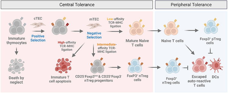
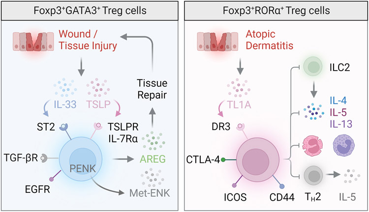
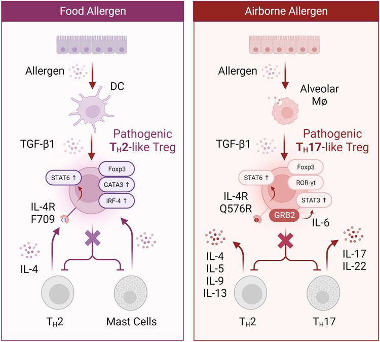
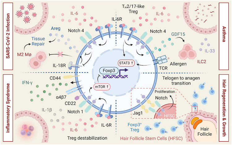
Regulatory T cells expressing the transcription factor forkhead box protein 3 mediate peripheral immune tolerance both to self-antigens and to the commensal flora. Their defective function due to inborn errors of immunity or acquired insults is associated with a broad range of autoimmune and immune dysregulatory diseases. Although their function in suppressing autoimmunity and enforcing commensalism is established, a broader role for regulatory T cells in tissue repair and metabolic regulation has emerged, enabled by unique programs of tissue adaptability and specialization. In this review, we focus on the myriad roles played by regulatory T cells in immunologic tolerance and host homeostasis and the potential to harness these cells in novel therapeutic approaches to human diseases.

Keywords: Foxp3; IL-2; Regulatory T cells; autoimmunity; immunologic tolerance; regulatory T-cell therapy.


Conflict of interest statement

Declaration of interests

S.H. has consulted for Merck KGaA. P.G. has consulted for RA Capital and Astro Therapeutics. A.H.S. currently has funding from Quark and AbbVie, unrelated to the submitted work. A.H.S. serves on advisory boards for SQZ Biotechnologies, Selecta, Elpiscience, Monopteros, Bicara, Fibrogen, IOME, Corner Therapeutics and Alixia. She also is on scientific advisory boards for the Massachusetts General Cancer Center, Program in Cellular and Molecular Medicine at Boston Children’s Hospital, the Human Oncology and Pathogenesis Program at Memorial Sloan Kettering Cancer Center, Bloomberg-Kimmel Institute for Cancer Immunotherapy, Glaxo Smith Kline, Janssen and Amgen. She is an academic editor for the Journal of Experimental Medicine. A.H.S. has patents/pending royalties on the PD-1 pathway from Roche and Novartis. M.C.H. has patents pending on the PHD3 pathway, is on the scientific advisory board for Alixia, Minovia, and MitoQ, and had research funding from Roche.

Figures

Figure 1
Figure 1. Central and peripheral immunological tolerance
Although T cells originate from the fetal liver and adult bone marrow, the thymus is the major site of T cell maturation. The selection of developing T cells is dependent on T cell receptor (TCR) recognition of self-peptide-major histocompatibility complex (self-peptide-MHC) molecules and results in the elimination of potentially harmful T cell clones. Positive selection is a process mediated by cortical thymic epithelial cells (cTEC) during which immature thymocytes whose TCRs fail to recognize self-peptide-MHC undergo a default pathway of apoptosis; a phenomenon known as “death by neglect”. Thymocytes whose TCRs recognize peptide-MHC presented by medullary thymic epithelial cells (mTEC) with high avidity undergo a process of negative selection resulting in apoptosis. In an alternative process that is incompletely understood, thymocytes bearing TCRs with intermediate avidities towards self-peptide-MHC are selected onto the Treg cell lineage via at least two distinct progenitor subsets including a CD25+Foxp3 and CD25 Foxp3low regulatory T cell population. The elimination of potentially harmful self-reactive T cells in the thymus through negative selection is the basis for0 central tolerance. In contrast, peripheral tolerance is induced in mature lymphocytes by self-antigens, including components of the extended self and innocuous environmental antigens by a pool of regulatory T cells derived from the thymus (nTreg) or generated in the periphery (pTreg) that act in concert to maintain organismal-wide immunological tolerance.
Figure 2
Figure 2. Developmental trajectory and subsets of regulatory T cells
The regulatory T cell pool is composed of two major populations that act in concert to maintain peripheral tolerance. Natural regulatory T (nTreg) cells develop in the thymus and play a critical role in the maintenance of tolerance to self-antigens. Peripheral regulatory T (pTreg) cells are generated from conventional naïve CD4+ T cells at mucosal interfaces and barrier sites and are critically important for the maintenance of tolerance towards dietary, environmental and commensal antigens. Regulatory T cells can also be induced (iTreg) in-vitro from conventional naïve CD4+ T cells following TCR ligation in the presence of transforming growth factor beta (TGF-β). Although all three populations express high levels of the transcription factor Foxp3 and share the expression of several canonical Treg cell surface molecules including CTLA-4, CD25 and GITR, the TCR repertoires of nTreg and pTreg/iTreg and are distinct and non-overlapping. Although not uniquely expressed by nTreg cells, the Ikaros family transcription factor Helios and cell surface glycoprotein neuropilin-1 (Nrp-1) are highly expressed in nTreg as opposed to pTreg/iTreg populations. pTreg/iTreg cells exhibit a greater susceptibility towards destabilization and loss of Foxp3 expression, largely due to the absence of an nTreg-like specific demethylation signature at conserved non-coding sequences within the locus of Foxp3 and other Treg signature genes.
Figure 3
Figure 3. Regulatory T cells in the skin
The skin Treg cell compartment is largely composed of nTreg cells that express the transcription factor GATA-3 (left), although a RORα+ population involved in the control of cutaneous inflammation has also been reported (right). These RORα+ skin Treg cells express the tumor necrosis factor receptor superfamily member 25, also known as death receptor 3 (DR3), and sequester TNF ligand–related molecule 1 (TL1A) to curtail type 2 innate lymphoid cell activity, eosinophil and basophil accumulation, and type 2 inflammation during atopic dermatitis. GATA-3+ skin Treg cells expressing the transcription factor IRF4 play a key role in skin wound repair and healing. Expression of alarmin receptors including the IL-33 receptor (ST2), IL-18 receptor and thymic stromal lymphopoietin receptor (TSLPR) enable skin Treg cells to sense and respond to cues associated with cutaneous damage and stress. Skin Treg cells also express the epidermal growth factor receptor (EGFR), which is thought to contribute to the accumulation of Treg cells in wounded skin and promote wound closure, likely though the production of the tissue reparative factor amphiregulin (AREG). Following UV light exposure, production of the opioid precursor proenkephalin (PENK) by UVB-exposed skin Treg cells contributes to generation of the neuropeptide methionine enkephalin (Met-ENK), which in turn promotes keratinocyte outgrowth and wound healing.
Figure 4
Figure 4. Pathological reprogramming of regulatory T cells in the intestine and lung
Left: Food allergic Il4raF709 mice exhibit deficits in allergen-specific Treg cells. OVA-specific Il4raF709 Treg cells fail to suppress food allergy as a result of undergoing pathogenic TH2 cell-like reprogramming due to excessive IL-4R-STAT6 signaling. These pathogenic TH2 cell-like Tregs are characterized by heightened expression of canonical TH2 program components such as IRF4 and GATA-3. Importantly, allergen-specific Treg cells of human subjects with food allergy harbor similar TH2 cell-like Tregs, suggesting that blockade of the IL-4R pathway may offer a viable therapeutic strategy for the induction of long-lasting tolerance. Right: Destabilization of IL-4raQ576R pTreg cells in asthma towards a pathogenic TH17 cell-like state leads to the exacerbation of pulmonary inflammation. IL-4raQ576-dependent recruitment of the growth-factor-receptor-bound protein 2 (GRB2) results in mitogen-activated protein kinase (MAPK) activation and heightened autocrine IL-6-STAT3 signaling which collectively derails protective pTreg cell responses in the lung during allergic airway inflammation. Blocking the IL-4R or IL-6R pathways may prevent the destabilization of allergen-specific pTreg cells into TH2 and TH17 like cells, respectively, and afford novel Treg intervention strategies for the re-establishment of tolerance.
Figure 5
Figure 5. Multifaced roles of Notch signaling in regulatory T cells
Notch signaling plays pleiotropic roles within the Treg cell compartment and is critically involved in the control of Treg cell function in the periphery. Upper Right: In experimental models of asthma, Notch4 expression on lung pTreg cells licenses allergic airway inflammation through a process that involves the destabilization of pTreg cells towards TH2 and TH17 cell fates. During allergic airway inflammation, Notch4 expression on lung pTreg cells also potentiates ILC2 activation and expansion, via a growth and differentiation factor 15 (GDF15)dependent mechanism that abrogates pTreg cell-mediated suppression of ILC2s. Upper Left: During severe SARS-CoV-2 infection, pTreg cell Notch4 represses production of the tissue reparative factor amphiregulin (AREG) by inhibiting IL-18 production, which promotes severe lung inflammation. Lower Left: In multisystem inflammatory syndrome in children (MIS-C), subjects harboring mutations in the Notch-related genes, upregulation of Notch1 on Treg cells leads to super-induction of CD22 and augmented TCR signaling, collectively promoting Treg-cell destabilization in an mTOR-dependent manner. Lower Right: Expression of the Notch ligand Jagged-1 by skin Treg cells promotes the proliferation and function of hair follicle stem cells to support hair follicle regeneration and hair growth.
Figure 6
Figure 6. Regulatory T cell therapies
A number of therapeutic approaches are currently attempting to harness the potential of Treg cells as therapeutics for autoimmune and immune dysregulatory disorders. These strategies include: A. genomic engineering strategies to artificially induce long-lasting Foxp3 expression and reprogram conventional CD4+ T cells into Foxp3-expressing Treg-like cells capable of inducing tolerance. B. Modified IL-2 reagents including IL-2 muteins, pegylated-IL-2 variants, IL-2:anti-IL-2 complexes and fusion proteins as well as orthogonal IL-2-IL-2R pairs. C. Adoptive cell therapies (ACTs) utilizing autologous ex-vivo expended polyclonal or antigen-specific Treg cells with engineered TCRs including chimeric antigen receptor (CAR) Treg cells.

References

    1. Silverstein AM. Autoimmunity versus horror autotoxicus: the struggle for recognition. Nat Immunol 2001; 2:279–81. - PubMed
    1. Brent L. The discovery of immunologic tolerance. Hum Immunol 1997; 52:75–81. - PubMed
    1. Kappler JW, Roehm N, Marrack P. T cell tolerance by clonal elimination in the thymus. Cell 1987; 49:273–80. - PubMed
    1. Kisielow P, Bluthmann H, Staerz UD, Steinmetz M, von Boehmer H. Tolerance in T-cell-receptor transgenic mice involves deletion of nonmature CD4+8+ thymocytes. Nature 1988; 333:742–6. - PubMed
    1. Anderson MS, Venanzi ES, Klein L, Chen Z, Berzins SP, Turley SJ, et al. Projection of an immunological self shadow within the thymus by the aire protein. Science 2002; 298:1395–401. - PubMed

Publication types

Substances