. 2023 Dec 8;69(6):308-316.
doi: 10.1262/jrd.2023-023. Epub 2023 Sep 30.

Fluctuation of CD9/SOX2-positive cell populations during the turnover of GH- and TSH-producing cells in the adult anterior pituitary gland

Affiliations

Fluctuation of CD9/SOX2-positive cell populations during the turnover of GH- and TSH-producing cells in the adult anterior pituitary gland

Kotaro Horiguchi et al. J Reprod Dev. .

Abstract

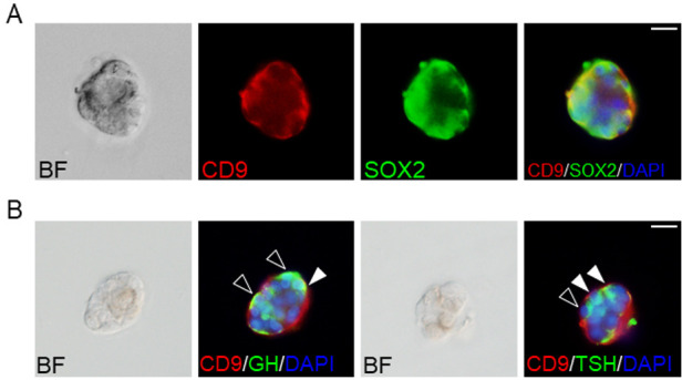
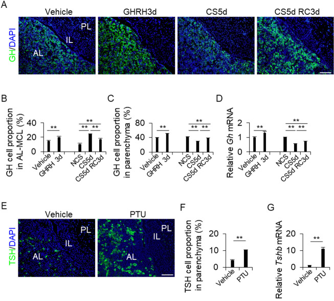
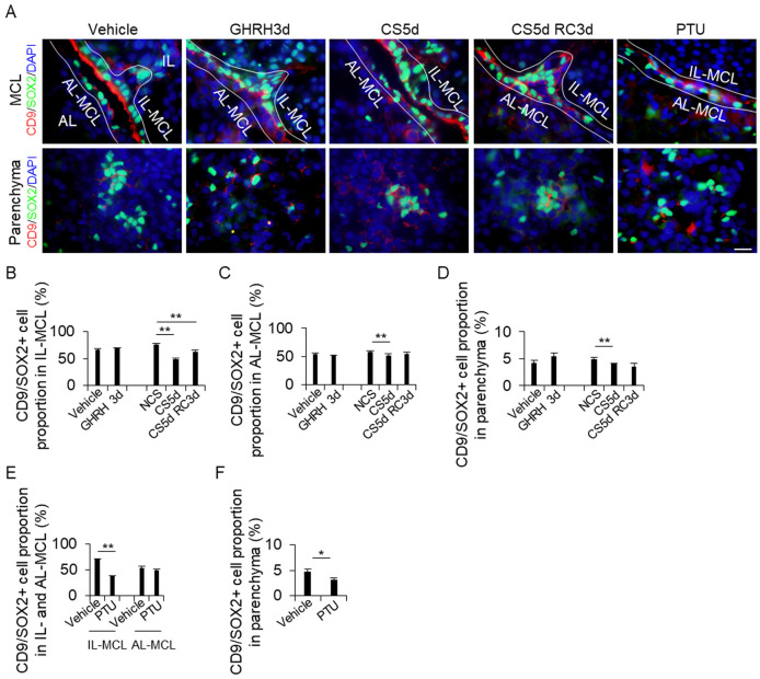
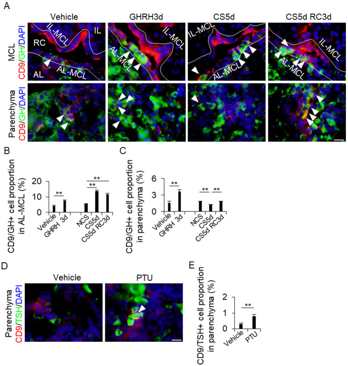
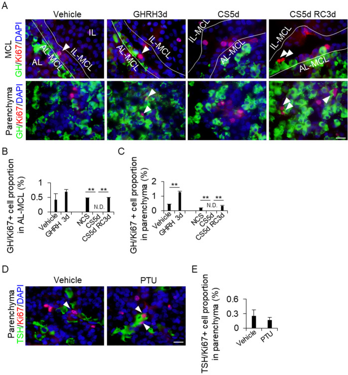
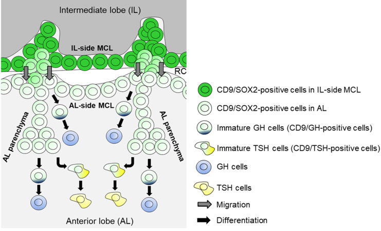
The adenohypophysis is comprised of the anterior and intermediate lobes (AL and IL, respectively). Cluster of differentiation 9 (CD9)- and sex-determining region Y-box 2 (SOX2)-positive cells are stem/progenitor hormone-producing cells in the AL. They are located in the marginal cell layer (MCL) facing Rathke's cleft between the AL and IL (primary niche) and the parenchyma of the AL (secondary niche). We previously showed that, in rats, CD9/SOX2-positive cells in the IL side of the MCL (IL-side MCL) migrate to the AL side (AL-side MCL) and differentiate into prolactin-producing cells (PRL cells) in the AL parenchyma during pregnancy, lactation, and diethylstilbestrol treatment, all of which increase PRL cell turnover. This study examined the changes in CD9/SOX2-positive stem/progenitor cell niches and their proportions by manipulating the turnover of growth hormone (GH)- and thyroid-stimulating hormone (TSH)-producing cells (GH and TSH cells, respectively), which are Pit1 lineage cells, as well as PRL cells. After induction, the isolated CD9/SOX2-positive cells from the IL-side MCL formed spheres and differentiated into GH and TSH cells. We also observed an increased GH cell proportion upon treatment with GH-releasing hormone and recovery from continuous stress and an increased TSH cell proportion upon propylthiouracil treatment, concomitant with alterations in the proportion of CD9/SOX2-positive cells in the primary and secondary niches. These findings suggest that CD9/SOX2-positive cells have the potential to supply GH and TSH when an increase in GH and TSH cell populations is required in the adult pituitary gland.

Keywords: Cluster of differentiation 9 (CD9); Growth hormone (GH); Pituitary; Sex-determining region Y-box 2 (SOX2); Thyroid-stimulating hormone (TSH).


Conflict of interest statement

The authors have nothing to declare.

Figures

Fig. 1.
Fig. 1.
Pituispheres of CD9/SOX2-positive cells in the intermediate lobes (IL) side of the marginal cell layer (MCL). (A) Double immunofluorescence staining for CD9 (red) and SOX2 (green) using CD9-positive pituispheres. The left panel shows a bright field (BF) image of the pituisphere. The right panel shows the merged image with DAPI (blue). (B) Hormone-producing cells in the CD9-positive pituispheres after induction. BF images and merged images with DAPI (blue), CD9 (red), and GH (green), or TSH (green) are shown. Black and white arrowheads indicate GH or TSH single-positive cells and CD9/GH or CD9/TSH double-positive cells, respectively. Scale bars: 20 μm (A and B).
Fig. 2.
Fig. 2.
(A and E) Immunohistochemistry for GH (A) and TSH (E) in the vehicle, GHRH treatment for 3 d (GHRH3d), continuous stress for 5 days (CS5d), and recovery for 3 days after CS for 5 days (CS5d RC3d). DAPI (blue) was added to identify the nuclei. (B, C, and F) Proportions of GH cells or TSH cells from the DAPI-positive cells in each treatment. The cells were counted in the AL-side MCL (AL-MCL) and the AL parenchyma (Parenchyma). NCS: no continuous stress, PTU: 2-week propylthiouracil treatment. (D and G) mRNA levels of Gh (D) and Tshb (G) in the AL in each treatment (mean ± SEM, n = 5). Data were normalized to that of Hprt1. The Gh and Tshb mRNA levels were calculated as the ratios of the NCS and vehicle values. PL, posterior lobe; IL, intermediate lobe; AL, anterior lobe. Scale bars: 100 μm (A and E). ** P < 0.01.
Fig. 3.
Fig. 3.
Detection of CD9/SOX2-positive cells in the IL-side and AL-side MCL and the AL parenchyma. (A) Double immunohistochemistry for CD9 and SOX2 in the vehicle, GH treatment for 3 d (GHRH3d), continuous stress for 5 days (CS5d), recovery for 3 days after CS for 5 days (CS5d RC3d), and 2-week propylthiouracil (PTU) treatment. The first and second rows show the MCL and the parenchyma, respectively. DAPI (blue) was added to identify the nuclei. (B) Proportion of CD9/SOX2-positive cells from the DAPI-positive cells in each treatment. The cells were counted in the IL-side MCL (IL-MCL; B and E), AL-side MCL (AL-MCL; C and E), and AL parenchyma (Parenchyma; D and F). AL, anterior lobe; IL, intermediate lobe; MCL, marginal cell layer. Scale bars: 20 μm (A). ** P < 0.01. * P < 0.05.
Fig. 4.
Fig. 4.
Detection of CD9/GH- or CD9/TSH-positive cells in the AL-side MCL and the AL parenchyma. (A and D) Double immunohistochemistry for CD9 (red) and GH (green) in the vehicle, GH treatment for 3 days (GHRH3d), continuous stress for 5 days (CS5d), and recovery for 3 days after CS for 5 days (CS5d RC3d) (A), and for CD9 (red) and TSH (green) in the vehicle and 2-week propylthiouracil treatments (PTU) (D). DAPI (blue) was added to identify the nuclei. White arrowheads indicate CD9/GH- (A) or CD9/TSH- (D) positive cells. (B, C, and E) Proportion of CD9/GH- (B and C) or CD9/TSH-positive cells (E) from the DAPI-positive cells in each treatment. The cells were counted in the AL-side MCL (AL-MCL; B) and AL parenchyma (Parenchyma; C and E). AL, anterior lobe; IL, intermediate lobe; MCL, marginal cell layer; PL, posterior lobe; RC, Rathke’s cleft; IL-MCL, IL side of the MCL. Scale bars: 20 μm (right panel of A and D). ** P < 0.01.
Fig. 5.
Fig. 5.
Proportion of GH/Ki67-positive cells in the IL-side and AL-side MCL and the AL parenchyma. (A and D) Double immunohistochemistry for GH (green) and Ki67 (red) in the vehicle, GH treatment for 3 days (GHRH3d), continuous stress for 5 days (CS5d), and recovery for 3 days after CS for 5 days (CS5d RC3d) (A), and for TSH (green) and Ki67 (red) in the vehicle and 2-week propylthiouracil treatments (PTU) (D). DAPI (blue) was added to identify the nuclei. White arrowheads indicate GH/Ki67- (A) or TSH/Ki67-positive cells (D). (B, C and E) Proportion of GH/Ki67-positive cells from the DAPI-positive cells in each treatment. The cells were counted in the AL-side MCL (AL-MCL; B) and AL parenchyma (Parenchyma; C and E). AL, anterior lobe; IL, intermediate lobe; MCL, marginal cell layer; IL-MCL, IL side of the MCL. Scale bars: 20 μm (A and B). ** P < 0.01.
Fig. 6.
Fig. 6.
Schematic of GH or TSH cell turnover in the adult AL and the supply of CD9/SOX2-positive stem cells. CD9/SOX2-positive cells in the IL-side MCL migrate into the AL-side MCL and then into the AL parenchyma. GH cells are derived from CD9/SOX2-positive stem cells in the AL-side MCL and the AL parenchyma. TSH cells are derived from CD9/SOX2-positive stem cells in the AL parenchyma. RC, Rathke’s cleft.

Similar articles

Cited by

References

    1. Kato Y, Yoshida S, Kato T. New insights into the role and origin of pituitary S100β-positive cells. Cell Tissue Res 2021; 386: 227–237. - PubMed
    1. Vila-Porcile E. The network of the folliculo-stellate cells and the follicles of the adenohypophysis in the rat (pars distalis). Z Zellforsch Mikrosk Anat 1972; 129: 328–369 (in French). - PubMed
    1. Fauquier T, Rizzoti K, Dattani M, Lovell-Badge R, Robinson IC. SOX2-expressing progenitor cells generate all of the major cell types in the adult mouse pituitary gland. Proc Natl Acad Sci USA 2008; 105: 2907–2912. - PMC - PubMed
    1. Horiguchi K, Fujiwara K, Takeda Y, Nakakura T, Tsukada T, Yoshida S, Hasegawa R, Takigami S, Ohsako S. CD9-positive cells in the intermediate lobe of the pituitary gland are important supplier for prolactin-producing cells in the anterior lobe. Cell Tissue Res 2021; 385: 713–726. - PubMed
    1. Horiguchi K, Fujiwara K, Yoshida S, Nakakura T, Arae K, Tsukada T, Hasegawa R, Takigami S, Ohsako S, Yashiro T, Kato T, Kato Y. Isolation and characterisation of CD9-positive pituitary adult stem/progenitor cells in rats. Sci Rep 2018; 8: 5533. - PMC - PubMed