Review
. 2023 Oct;55(10):2105-2115.
doi: 10.1038/s12276-023-01093-y. Epub 2023 Oct 2.

Spatial transcriptomics in neuroscience

Affiliations
Review

Spatial transcriptomics in neuroscience

Namyoung Jung et al. Exp Mol Med. 2023 Oct.

Abstract

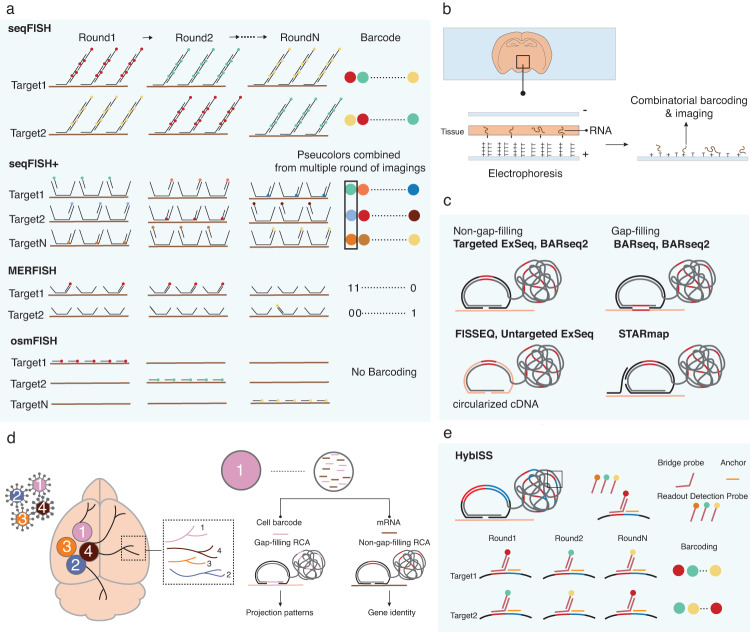
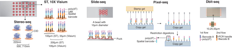
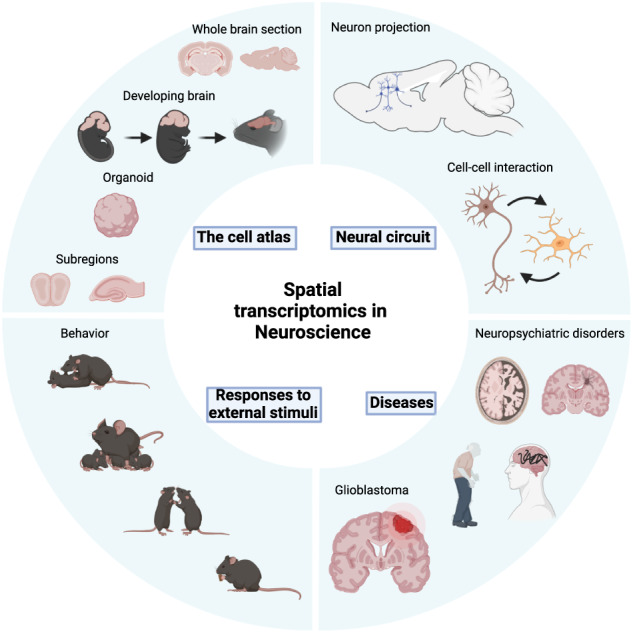
The brain is one of the most complex living tissue types and is composed of an exceptional diversity of cell types displaying unique functional connectivity. Single-cell RNA sequencing (scRNA-seq) can be used to efficiently map the molecular identities of the various cell types in the brain by providing the transcriptomic profiles of individual cells isolated from the tissue. However, the lack of spatial context in scRNA-seq prevents a comprehensive understanding of how different configurations of cell types give rise to specific functions in individual brain regions and how each distinct cell is connected to form a functional unit. To understand how the various cell types contribute to specific brain functions, it is crucial to correlate the identities of individual cells obtained through scRNA-seq with their spatial information in intact tissue. Spatial transcriptomics (ST) can resolve the complex spatial organization of cell types in the brain and their connectivity. Various ST tools developed during the past decade based on imaging and sequencing technology have permitted the creation of functional atlases of the brain and have pulled the properties of neural circuits into ever-sharper focus. In this review, we present a summary of several ST tools and their applications in neuroscience and discuss the unprecedented insights these tools have made possible.


Conflict of interest statement

The authors declare no competing interests.

Figures

Fig. 1
Fig. 1. An overview of imaging-based spatial transcriptomics (ST) technologies used in neuroscience.
a A schematic illustration of in situ hybridization (ISH)-based tools. Sequential FISH (seqFISH) identifies an RNA target by different combinations of fluorescent probes. An RNA is labeled with one color in each round of hybridization, enabling the colorimetric barcoding scheme. SeqFISH+ collapses multiple imaging rounds to generate ‘pseudocolor’ barcoding, in which an RNA is fluorescent in one of the sets of images and collapsed into one image corresponding to one round. Multiplexed error-robust FISH (MERFISH) exploits a combinatorial barcoding scheme in which a target RNA is assigned to a unique binary barcode, determined by on-off fluorescence in sequential rounds of hybridization. Cyclic-ouroboros FISH (osmFISH) does not use the combinatorial barcoding scheme but relies on multiple rounds of hybridization to label a target with a specific color in the image. b Enhanced electric FISH (EEL FISH). A tissue section is mounted on a capture slide coated with an electrically conductive layer of indium tin oxide (ITO). The ITO layer is modified with poly(D-lysine) and oligo(dT) to capture RNA electronically and chemically. The captured RNA is encoded and decoded by a combinatorial barcoding scheme. c A schematic illustration of the in situ sequencing (ISS)-based method. ISS-based techniques adopt various versions of padlock probes. Targeted expansion sequencing (ExSeq) and barcoded anatomy resolved by sequencing 2 (BARseq2) use non-gap-filling padlock probes, each of which contains a unique barcode. On the other hand, the gap-filling padlock probe used by BARseq and BARseq2 generates a barcode sequence complementary to a segment of the target sequence by gap-filling. Fluorescence in situ sequencing (FISSEQ) and untargeted ExSeq circularize cDNA, conferring padlock probe-like function on the cDNA. Spatially resolved transcript amplicon readout mapping (STARmap) uses SNAIL, in which a primer is hybridized to a target and provides a backbone to a padlock probe. The probes are amplified by rolling-circle amplification (RCA), generating a rolony containing a unique barcode for a target. In ISS-based technologies, fluorescent barcodes are generated by the incorporation of a fluorescent-conjugated nucleotide (sequencing-by-synthesis) or the ligation of fluorescent-conjugated oligonucleotides (sequencing-by-ligation). d BARseq2. BARseq2 can detect endogenous RNA expression and identify the projection pattern of the infected cell simultaneously. The neurons are barcoded by a viral infection, enabling the identification of projection patterns in different areas of the brain. The RNA barcode in a neuron is reverse transcribed and amplified by the gap-filling method. After amplification, the rolony containing the barcodes is sequenced to decipher neuron identity. The non-gap-filling approach is used for target gene detection. e Hybridization-based in situ sequencing (HybISS). HybISS integrates the advantages of ISH and ISS-based methods. HybISS uses the padlock probe and RCA to generate a rolony barcoded and decoded by a colorimetric scheme.
Fig. 2
Fig. 2. A schematic illustration of sequencing-based technologies.
In the original ST, capture probes containing poly(dT), unique molecular identifier (UMI), and a spatial barcode are printed in an array of spots with 100 μm in diameter. The ST is commercialized as Visium by 10X Genomics with a resolution of 55 μm in diameter. In spatiotemporal enhanced resolution omics sequencing (Stereo-seq), a DNA nanoball including a spatial barcode is printed on the slide as a 220 nm diameter spot, providing the highest resolution among sequencing-based techniques. Slide-seq uses a bead with a 10 μm diameter instead of a spot, forming a layer called a “puck” on a slide. Polony-indexed library sequencing (Pixel-seq) generates capture-probe printed gels with the same arrangements of the probes across different gels, hence greatly reducing the time and cost of producing and decoding spatially barcoded arrays. Deterministic barcoding in tissue for spatial omics sequencing (DBiT-seq) enables the simultaneous mapping of proteins and RNA. The capture probes are delivered to tissue by a microfluidic device in two flows. The first flow contains probes with poly(dT) and spatial barcode A, and the second flow delivers another probe containing spatial barcode B perpendicular to the first flow. The two barcodes A and B are joined at the intersection of the two flows, which forms a pixel with a unique combination of barcodes A and B. The resolution of DBiT-seq is 10, 25, and 50 μm, determined by the channel width of a microfluidic device.
Fig. 3
Fig. 3. The application of ST in neuroscience.
In neuroscience, ST has been employed to generate a cell atlas of various brain regions and has also been used to map neural circuits, to investigate the molecular and cellular responses to external stimuli and to characterize the differences between normal and diseased brains. (Created with BioRender.com).

Similar articles

Cited by

References

    1. Molnar Z, et al. New insights into the development of the human cerebral cortex. J. Anat. 2019;235:432–451. doi: 10.1111/joa.13055. - DOI - PMC - PubMed
    1. Le Tissier P, et al. An updated view of hypothalamic-vascular-pituitary unit function and plasticity. Nat. Rev. Endocrinol. 2017;13:257–267. doi: 10.1038/nrendo.2016.193. - DOI - PubMed
    1. Hodge RD, et al. Conserved cell types with divergent features in human versus mouse cortex. Nature. 2019;573:61–68. doi: 10.1038/s41586-019-1506-7. - DOI - PMC - PubMed
    1. Tasic B, et al. Adult mouse cortical cell taxonomy revealed by single cell transcriptomics. Nat. Neurosci. 2016;19:335–346. doi: 10.1038/nn.4216. - DOI - PMC - PubMed
    1. Tasic B, et al. Shared and distinct transcriptomic cell types across neocortical areas. Nature. 2018;563:72–78. doi: 10.1038/s41586-018-0654-5. - DOI - PMC - PubMed

Publication types