Review
. 2023 Oct;55(10):2147-2161.
doi: 10.1038/s12276-023-01101-1. Epub 2023 Oct 2.

A stepwise activation model for the insulin receptor

Affiliations
Review

A stepwise activation model for the insulin receptor

Na-Oh Yunn et al. Exp Mol Med. 2023 Oct.

Abstract

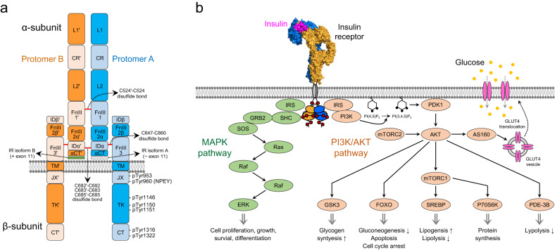
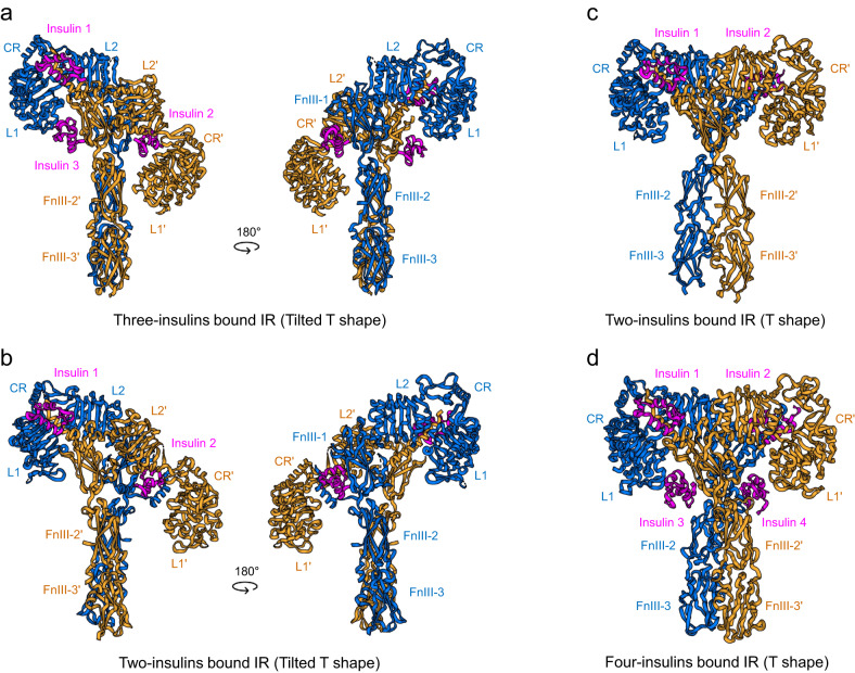
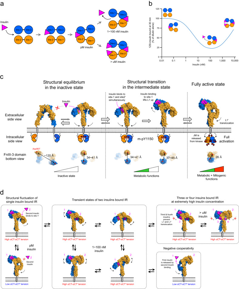
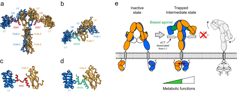
The binding of insulin to the insulin receptor (IR) triggers a cascade of receptor conformational changes and autophosphorylation, leading to the activation of metabolic and mitogenic pathways. Recent advances in the structural and functional analyses of IR have revealed the conformations of the extracellular domains of the IR in inactive and fully activated states. However, the early activation mechanisms of this receptor remain poorly understood. The structures of partially activated IR in complex with aptamers provide clues for understanding the initial activation mechanism. In this review, we discuss the structural and functional features of IR complexed with various ligands and propose a model to explain the sequential activation mechanism. Moreover, we discuss the structures of IR complexed with biased agonists that selectively activate metabolic pathways and provide insights into the design of selective agonists and their clinical implications.


Conflict of interest statement

The authors declare no competing interests.

Figures

Fig. 1
Fig. 1. Domain architecture and signal pathway of IR.
a IR domain architecture composed of two α-subunits and two β-subunits. Protomer A and B are colored blue and orange, respectively. A prime (′) symbol represents the opposite protomer element. Red lines: disulfide bonds. b Two major signaling pathways initiated from insulin-induced IR activation. Insulin binding to IR induces autophosphorylation on multiple tyrosine residues, which recruits and phosphorylates IRS and SHC. IRS proteins initiate the PI3K–AKT pathway by activating PI3K. SHC proteins mainly initiate the Grb2-SosRas-Raf-ERK cascade (MAPK pathway).
Fig. 2
Fig. 2. Inactive conformation and two insulin binding sites of IR.
a Schematic diagram of IR based on the apo (unliganded) structure. Protomer A and B are colored blue and orange, respectively. b The X-ray crystal structure of apo IR (PDB ID: 4ZXB). Two hook-like protomers form a symmetric Λ-shaped dimer in an antiparallel manner. c Orthogonal view of one-half of the Λ-shaped IR homodimer. Two insulin binding sites, site-1 and site-2’, are shown in red dotted circles. d Close-up view of insulin-bound site-1 in the Γ-shaped IR structure (PDB ID: 7YQ3). e Close-up view of insulin-bound site-2’ in the T-shaped IR (PDB: 6PXV). f Close-up view of the insulin bridge between site-1 and site-2’ in the two-insulin-bound tilted T-shaped structure (PDB ID: 7STK). g Amino acid sequence and structure (PDB ID: 1MSO) of human insulin. The A-chain and B-chain are colored green and cyan, respectively. Yellow lines indicate disulfide bonds. h Superimposition of free insulin (PDB ID: 1MSO; green and cyan) and insulin-bound site-1 (PDB ID: 6VEP; pink). i Superimposition of free insulin (PDB ID: 1MSO; green and cyan) and insulin-bound site-2’ (PDB ID: 6SOF; pink).
Fig. 3
Fig. 3. Fully active Г-shaped IR bound to a single insulin molecule.
a Cryo-EM structure of the IR extracellular region with the leucine-zipper domain fused to the C-terminus (6HN5 and 6HN4). b Cryo-EM structure of full-length IR bound to one insulin molecule and one A43 aptamer (PDB ID: 7YQ3). The A43 aptamer fits in a pocket comprising FnIII-1, CR′, and L2′ on the insulin-free side. c Close-up view of αCT and αCT′ interacting with L1’ and L1, respectively, in the apo IR structure (PDB ID: 4ZXB). The crosslink between insert domains is missing in the structure. d Close-up view of the αCT-αCT′ bridge cross-linked by the C683-C683’ disulfide bond in the A43-bound Γ-shaped IR structure. e A model for the insulin-enhancing activity of the A43 aptamer. When a single insulin molecule binds to IR leads to the Γ-shaped conformation, the position of the opposite L1’ may become unstable. Because of the αCT-αCT′ bridge, the structural instability of L1’ can interfere with insulin binding to site-1 (L1’ and αCT′) by increasing steric tension between L1 and L1′. A43 binding to IR can stabilize the Γ-shaped conformation by fixing the position of L1’, which enhances insulin-induced IR phosphorylation by stabilizing insulin binding to site-1. f Close-up view of A43 in a pocket comprising FnIII-1, CR′, and L2′ in Fig. 3b. g Superposition of insulin bound to site-1 in the Γ-shaped IR structure onto the Λ-shaped apo IR by aligning the L1 domain to demonstrate the steric clash of insulin. Steric clash of insulin (pink) with FnIII-1’ and FnIII-2’ (orange) is evident.
Fig. 4
Fig. 4. A62 aptamer-trapped monophosphorylated IR.
a The cryo-EM structure of full-length IR bound to two A62 aptamers (PDB ID: 7YQ6). The A62 aptamers are green. b Superposition of the protomers of the Λ-shaped apo structure (green, Fig. 2b) and arrowhead-shaped IR structure (red, Fig. 4a). c Comparison of the Λ-shaped apo IR structures (left) and two A62-bound IR structures (right). Protomer rotation by 26.7° with respect to Cys524 converts the Λ-shaped IR structure to the arrowhead-shaped IR structure. d Close-up view of the A62 bridge established between L1 and FnIII-1′ in arrowhead-shaped IR. e Superimposition of insulin-bound site-1 (PDB ID: 7YQ3) and A62-bound L1 in arrowhead-shaped IR. f Superimposition of insulin-bound site-2’ (PDB ID: 6PXV) and A62-bound FnIII-1’ in the arrowhead-shaped IR. The A62 binding site overlaps with insulin-binding site-1 and site-2’. g The cryo-EM structure of the tilted T-shaped IR complexed with one A62 aptamer and one insulin molecule (PDB ID: 7YQ5) in three different views.
Fig. 5
Fig. 5. The cryo-EM structures of IR bound with two, three or four insulin molecules.
The structure of tilted T-shaped IR complexed with three human–venom insulin hybrids (PDB ID: 7PG3) a with two insulin molecules (PDB ID: 7STK). b The cryo-EM structure of T-shaped IR bound to two insulin molecules (PDB ID: 8GUY) and c with three insulin molecules (PDB ID: 6SOF). d Insulin and insulin analogs are pink.
Fig. 6
Fig. 6. The proposed model for insulin-induced IR activation and negative cooperativity of insulin binding.
a A conventional bivalent-crosslinking model showing the insulin binding mechanism,. At low insulin concentrations (~pM), the first insulin molecule binds strongly to the IR dimer by simultaneously interacting with site-1 and site-2’. As the insulin concentration increases (1–100 nM), the dissociation of previously bound insulin from IR is accelerated by a second insulin molecule binding to the opposite site-1’ and site-2 (negative cooperativity). At very high insulin concentrations (>μM), the binding of the second and third insulin molecule to each site-1’ and site-2 prevents the dissociation of the previously bound insulin from IR. b Dose‒response curve for the negative cooperativity of insulin binding to IR,. The bell-shaped curve indicates that accelerated dissociation of bound insulin changes in a concentration-dependent manner. c Stepwise activation model for insulin receptor activation from the extracellular and intracellular views. Insulin is pink, and arrows with dotted lines represent critical conformational changes in each step. The FnIII-3 domain bottom view represents the distance between the membrane proximal ends of FnIII-3 and FnIII-3′ in each step. d Model for the negative cooperativity of insulin binding. The dissociation of previously bound insulin is initiated by binding of a second insulin molecule to site-1’ and site-2. Two-insulin-bound IR undergoes transitions among various conformational states due to its intrinsic instability. When two insulin-bound IR structures transition into the Γ-shaped structure, previously bound insulin is dissociated from IR.
Fig. 7
Fig. 7. Functional selectivity of IR.
a The cryo-EM structure of extended T-shaped IR bound to two S597 (PDB ID: 8DTL). b The cryo-EM structure of partial domains of IR bound to IM459 (PDB ID: 7U6D). Detailed binding mode of S597 (c) and IM459 (d). Both peptides bind to site-1 and site-2’ simultaneously, similar to the A62 in the arrowhead-shaped IR, which forms a bridge. e Proposed model of the functional selectivity of IR. Cross-linking between L1 and FnIII-1’ with the dissociation of αCT′ from L1 establishes the intermediate state of IR, which may be critical for its metabolic effects.

References

    1. Haeusler RA, McGraw TE, Accili D. Biochemical and cellular properties of insulin receptor signalling. Nat. Rev. Mol. Cell Biol. 2018;19:31–44. doi: 10.1038/nrm.2017.89. - DOI - PMC - PubMed
    1. Belfiore, A. et al. Insulin receptor isoforms in physiology and disease: an updated view. Endocr. Rev. 38, 379–431 (2017). - PMC - PubMed
    1. Cai W, et al. Domain-dependent effects of insulin and IGF-1 receptors on signalling and gene expression. Nat. Commun. 2017;8:14892. doi: 10.1038/ncomms14892. - DOI - PMC - PubMed
    1. Worster DT, et al. Akt and ERK control the proliferative response of mammary epithelial cells to the growth factors IGF-1 and EGF through the cell cycle inhibitor p57Kip2. Sci. Signal. 2012;5:ra19. doi: 10.1126/scisignal.2001986. - DOI - PMC - PubMed
    1. Diagnosis and classification of diabetes mellitus. Diabetes Care37, S81-S90 (2014). - PubMed

Publication types

MeSH terms