

Automated detection of cardiac rest period for trigger delay calculation for image-based navigator coronary magnetic resonance angiography
- PMID: 37779192
- PMCID: PMC10544388
- DOI: 10.1186/s12968-023-00962-9
Automated detection of cardiac rest period for trigger delay calculation for image-based navigator coronary magnetic resonance angiography
Abstract
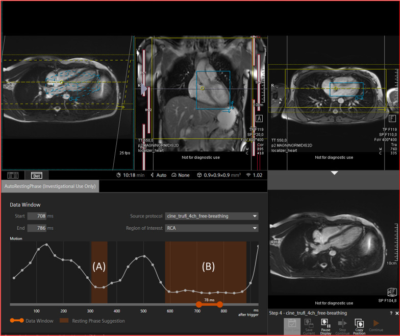
Background: Coronary magnetic resonance angiography (coronary MRA) is increasingly being considered as a clinically viable method to investigate coronary artery disease (CAD). Accurate determination of the trigger delay to place the acquisition window within the quiescent part of the cardiac cycle is critical for coronary MRA in order to reduce cardiac motion. This is currently reliant on operator-led decision making, which can negatively affect consistency of scan acquisition. Recently developed deep learning (DL) derived software may overcome these issues by automation of cardiac rest period detection.
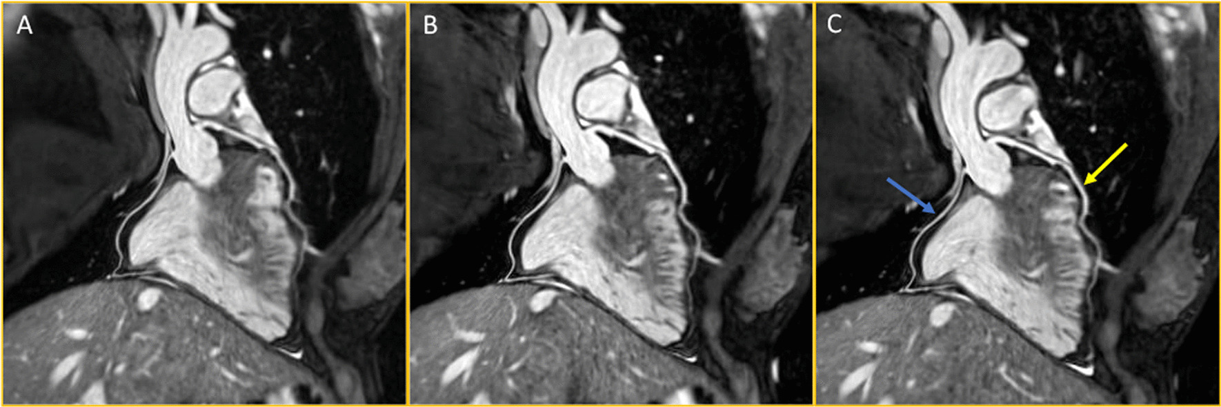
Methods: Thirty individuals (female, n = 10) were investigated using a 0.9 mm isotropic image-navigator (iNAV)-based motion-corrected coronary MRA sequence. Each individual was scanned three times utilising different strategies for determination of the optimal trigger delay: (1) the DL software, (2) an experienced operator decision, and (3) a previously utilised formula for determining the trigger delay. Methodologies were compared using custom-made analysis software to assess visible coronary vessel length and coronary vessel sharpness for the entire vessel length and the first 4 cm of each vessel.
Results: There was no difference in image quality between any of the methodologies for determination of the optimal trigger delay, as assessed by visible coronary vessel length, coronary vessel sharpness for each entire vessel and vessel sharpness for the first 4 cm of the left mainstem, left anterior descending or right coronary arteries. However, vessel length of the left circumflex was slightly greater using the formula method. The time taken to calculate the trigger delay was significantly lower for the DL-method as compared to the operator-led approach (106 ± 38.0 s vs 168 ± 39.2 s, p < 0.01, 95% CI of difference 25.5-98.1 s).
Conclusions: Deep learning-derived automated software can effectively and efficiently determine the optimal trigger delay for acquisition of coronary MRA and thus may simplify workflow and improve reproducibility.
Keywords: Cardiac magnetic resonance angiography; Cardiac rest period; Deep learning.
© 2023. Society for Cardiovascular Magnetic Resonance.
Conflict of interest statement
The authors declare that they have no competing interests.
Figures
Similar articles
-
Motion corrected water/fat whole-heart coronary MR angiography with 100% respiratory efficiency.Magn Reson Med. 2019 Aug;82(2):732-742. doi: 10.1002/mrm.27732. Epub 2019 Mar 29. Magn Reson Med. 2019. PMID: 30927310 Free PMC article.
-
Visualization of coronary arteries in paediatric patients using whole-heart coronary magnetic resonance angiography: comparison of image-navigation and the standard approach for respiratory motion compensation.J Cardiovasc Magn Reson. 2019 Feb 25;21(1):13. doi: 10.1186/s12968-019-0525-8. J Cardiovasc Magn Reson. 2019. PMID: 30798789 Free PMC article.
-
3D whole-heart isotropic sub-millimeter resolution coronary magnetic resonance angiography with non-rigid motion-compensated PROST.J Cardiovasc Magn Reson. 2020 Apr 16;22(1):24. doi: 10.1186/s12968-020-00611-5. J Cardiovasc Magn Reson. 2020. PMID: 32299445 Free PMC article.
-
Robust volume-targeted balanced steady-state free-precession coronary magnetic resonance angiography in a breathhold at 3.0 Tesla: a reproducibility study.J Cardiovasc Magn Reson. 2014 Apr 23;16(1):27. doi: 10.1186/1532-429X-16-27. J Cardiovasc Magn Reson. 2014. PMID: 24758168 Free PMC article.
-
Whole-heart non-rigid motion corrected coronary MRA with autofocus virtual 3D iNAV.Magn Reson Imaging. 2022 Apr;87:169-176. doi: 10.1016/j.mri.2022.01.007. Epub 2022 Jan 7. Magn Reson Imaging. 2022. PMID: 34999163
Cited by
-
High-resolution automated free-breathing coronary magnetic resonance angiography in comparison with coronary computed tomography angiography.Eur Heart J Imaging Methods Pract. 2025 Mar 27;3(1):qyaf037. doi: 10.1093/ehjimp/qyaf037. eCollection 2025 Jan. Eur Heart J Imaging Methods Pract. 2025. PMID: 40330538 Free PMC article.
-
A comprehensive evaluation of the left atrium using cardiovascular magnetic resonance.J Cardiovasc Magn Reson. 2025 Summer;27(1):101852. doi: 10.1016/j.jocmr.2025.101852. Epub 2025 Feb 5. J Cardiovasc Magn Reson. 2025. PMID: 39920924 Free PMC article. Review.
-
Highlights of the society for magnetic resonance angiography 2024 conference.J Cardiovasc Magn Reson. 2025 Summer;27(1):101878. doi: 10.1016/j.jocmr.2025.101878. Epub 2025 Mar 12. J Cardiovasc Magn Reson. 2025. PMID: 40086635 Free PMC article.
-
The beating heart: artificial intelligence for cardiovascular application in the clinic.MAGMA. 2024 Jul;37(3):369-382. doi: 10.1007/s10334-024-01180-9. Epub 2024 Jun 22. MAGMA. 2024. PMID: 38907767 Free PMC article. Review.
References
-
- Bhat H, Ge L, Nielles-Vallespin S, Zuehlsdorff S, Li D. 3D radial sampling and 3D affine transform-based respiratory motion correction technique for free-breathing whole-heart coronary MRA with 100% imaging efficiency. Magn Reson Med. 2011;65(5):1269–1277. doi: 10.1002/mrm.22717. - DOI - PMC - PubMed
Publication types
MeSH terms
Grants and funding
LinkOut - more resources
Full Text Sources
Miscellaneous
