

Meiotic and mitotic aneuploidies drive arrest of in vitro fertilized human preimplantation embryos
- PMID: 37779206
- PMCID: PMC10544495
- DOI: 10.1186/s13073-023-01231-1
Meiotic and mitotic aneuploidies drive arrest of in vitro fertilized human preimplantation embryos
Abstract
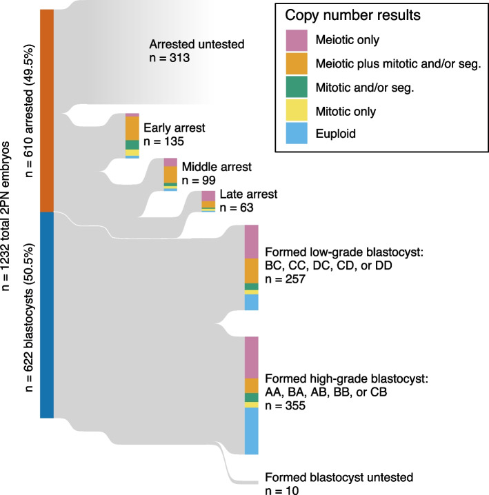
Background: The high incidence of aneuploidy in early human development, arising either from errors in meiosis or postzygotic mitosis, is the primary cause of pregnancy loss, miscarriage, and stillbirth following natural conception as well as in vitro fertilization (IVF). Preimplantation genetic testing for aneuploidy (PGT-A) has confirmed the prevalence of meiotic and mitotic aneuploidies among blastocyst-stage IVF embryos that are candidates for transfer. However, only about half of normally fertilized embryos develop to the blastocyst stage in vitro, while the others arrest at cleavage to late morula or early blastocyst stages.
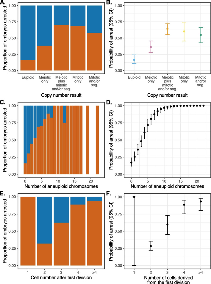
Methods: To achieve a more complete view of the impacts of aneuploidy, we applied low-coverage sequencing-based PGT-A to a large series (n = 909) of arrested embryos and trophectoderm biopsies. We then correlated observed aneuploidies with abnormalities of the first two cleavage divisions using time-lapse imaging (n = 843).
Results: The combined incidence of meiotic and mitotic aneuploidies was strongly associated with blastocyst morphological grading, with the proportion ranging from 20 to 90% for the highest to lowest grades, respectively. In contrast, the incidence of aneuploidy among arrested embryos was exceptionally high (94%), dominated by mitotic aneuploidies affecting multiple chromosomes. In turn, these mitotic aneuploidies were strongly associated with abnormal cleavage divisions, such that 51% of abnormally dividing embryos possessed mitotic aneuploidies compared to only 23% of normally dividing embryos.
Conclusions: We conclude that the combination of meiotic and mitotic aneuploidies drives arrest of human embryos in vitro, as development increasingly relies on embryonic gene expression at the blastocyst stage.
Keywords: IVF; Meiosis; Mitosis; Monosomy; Preimplantation genetic testing; Time-lapse; Trisomy.
© 2023. BioMed Central Ltd., part of Springer Nature.
Conflict of interest statement
RCM is co-inventor on a patent application by Johns Hopkins University related to inferring the origins of aneuploidies from PGT-A data. KA is the owner and scientific director of the London Women’s Clinic. AHH, MCS, CO, and AM declare no competing interests.
Figures


Similar articles
-
Preimplantation genetic testing for aneuploidies (abnormal number of chromosomes) in in vitro fertilisation.Cochrane Database Syst Rev. 2020 Sep 8;9(9):CD005291. doi: 10.1002/14651858.CD005291.pub3. Cochrane Database Syst Rev. 2020. PMID: 32898291 Free PMC article.
-
The incidence and origin of segmental aneuploidy in human oocytes and preimplantation embryos.Hum Reprod. 2017 Dec 1;32(12):2549-2560. doi: 10.1093/humrep/dex324. Hum Reprod. 2017. PMID: 29126206
-
Copy number analysis of meiotic and postzygotic mitotic aneuploidies in trophectoderm cells biopsied at the blastocyst stage and arrested embryos.Prenat Diagn. 2021 Apr;41(5):525-535. doi: 10.1002/pd.5816. Epub 2020 Sep 9. Prenat Diagn. 2021. PMID: 32833230 Review.
-
Incidence and origin of meiotic whole and segmental chromosomal aneuploidies detected by karyomapping.Reprod Biomed Online. 2019 Mar;38(3):330-339. doi: 10.1016/j.rbmo.2018.11.023. Epub 2018 Dec 23. Reprod Biomed Online. 2019. PMID: 30639160
-
The origin and significance of additional aneuploidy events in couples undergoing preimplantation genetic diagnosis for translocations by array comparative genomic hybridization.Reprod Biomed Online. 2016 Feb;32(2):178-89. doi: 10.1016/j.rbmo.2015.11.017. Epub 2015 Dec 2. Reprod Biomed Online. 2016. PMID: 26738467
Cited by
-
CHK1-CDC25A-CDK1 regulate cell cycle progression and protect genome integrity in early mouse embryos.EMBO Rep. 2023 Oct 9;24(10):e56530. doi: 10.15252/embr.202256530. Epub 2023 Sep 11. EMBO Rep. 2023. PMID: 37694680 Free PMC article.
-
Cell Competition Eliminates Aneuploid Human Pluripotent Stem Cells.bioRxiv [Preprint]. 2024 May 10:2024.05.08.593217. doi: 10.1101/2024.05.08.593217. bioRxiv. 2024. Update in: Stem Cell Reports. 2025 Jun 10;20(6):102506. doi: 10.1016/j.stemcr.2025.102506. PMID: 38766106 Free PMC article. Updated. Preprint.
-
Approximate Bayesian computation supports a high incidence of chromosomal mosaicism in blastocyst-stage human embryos.bioRxiv [Preprint]. 2024 Dec 2:2024.11.26.625484. doi: 10.1101/2024.11.26.625484. bioRxiv. 2024. Update in: Genetics. 2025 Aug 01:iyaf149. doi: 10.1093/genetics/iyaf149. PMID: 39677623 Free PMC article. Updated. Preprint.
-
Unraveling the mysteries of early embryonic arrest: genetic factors and molecular mechanisms.J Assist Reprod Genet. 2024 Dec;41(12):3301-3316. doi: 10.1007/s10815-024-03259-7. Epub 2024 Sep 26. J Assist Reprod Genet. 2024. PMID: 39325344 Review.
-
Biopsy vs comprehensive embryo/blastocyst analysis: a closer look at embryonic chromosome evaluation.Hum Reprod Open. 2025 Mar 12;2025(2):hoaf013. doi: 10.1093/hropen/hoaf013. eCollection 2025. Hum Reprod Open. 2025. PMID: 40123894 Free PMC article.
References
-
- Gardner RJM, Amor DJ. Gardner and Sutherland’s Chromosome Abnormalities and Genetic Counseling. Oxford: Oxford University Press; 2018.
-
- Levy B, Sigurjonsson S, Pettersen B, Maisenbacher MK, Hall MP, Demko Z, et al. Genomic imbalance in products of conception: single-nucleotide polymorphism chromosomal microarray analysis. Obstet Gynecol. 2014;124:202–209. - PubMed
-
- Segawa T, Kuroda T, Kato K, Kuroda M, Omi K, Miyauchi O, et al. Cytogenetic analysis of the retained products of conception after missed abortion following blastocyst transfer: a retrospective, large-scale, single-centre study. Reprod Biomed Online. 2017;34:203–210. - PubMed
Publication types
MeSH terms
Grants and funding
LinkOut - more resources
Full Text Sources
