

Theranostic in GLP-1R molecular imaging: challenges and emerging opportunities
- PMID: 37780209
- PMCID: PMC10540701
- DOI: 10.3389/fmolb.2023.1210347
Theranostic in GLP-1R molecular imaging: challenges and emerging opportunities
Abstract
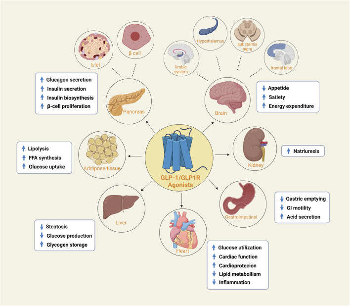
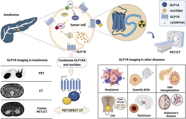
Theranostic in nuclear medicine combines diagnostic imaging and internal irradiation therapy using different therapeutic nuclear probes for visual diagnosis and precise treatment. GLP-1R is a popular receptor target in endocrine diseases, non-alcoholic steatohepatitis, tumors, and other areas. Likewise, it has also made breakthroughs in the development of molecular imaging. It was recognized that GLP-1R imaging originated from the study of insulinoma and afterwards was expanded in application including islet transplantation, pancreatic β-cell mass measurement, and ATP-dependent potassium channel-related endocrine diseases. Fortunately, GLP-1R molecular imaging has been involved in ischemic cardiomyocytes and neurodegenerative diseases. These signs illustrate the power of GLP-1R molecular imaging in the development of medicine. However, it is still limited to imaging diagnosis research in the current molecular imaging environment. The lack of molecular-targeted therapeutics related report hinders its radiology theranostic. In this article, the current research status, challenges, and emerging opportunities for GLP-1R molecular imaging are discussed in order to open a new path for theranostics and to promote the evolution of molecular medicine.
Keywords: GLP-1r; challenges; molecular imaging; opportunities; theranostics.
Copyright © 2023 Xie, Wang, Pei and Chen.
Conflict of interest statement
The authors declare that the research was conducted in the absence of any commercial or financial relationships that could be construed as a potential conflict of interest.
Figures
Similar articles
-
Theranostics in neuroendocrine tumors: an overview of current approaches and future challenges.Rev Endocr Metab Disord. 2021 Sep;22(3):581-594. doi: 10.1007/s11154-020-09552-x. Rev Endocr Metab Disord. 2021. PMID: 32495250 Review.
-
Development of an 111In-Labeled Glucagon-Like Peptide-1 Receptor-Targeting Exendin-4 Derivative that Exhibits Reduced Renal Uptake.Mol Pharm. 2022 Mar 7;19(3):1019-1027. doi: 10.1021/acs.molpharmaceut.2c00068. Epub 2022 Feb 9. Mol Pharm. 2022. PMID: 35138111
-
Cinchonine, a Potential Oral Small-Molecule Glucagon-Like Peptide-1 Receptor Agonist, Lowers Blood Glucose and Ameliorates Non-Alcoholic Steatohepatitis.Drug Des Devel Ther. 2023 May 11;17:1417-1432. doi: 10.2147/DDDT.S404055. eCollection 2023. Drug Des Devel Ther. 2023. PMID: 37197367 Free PMC article.
-
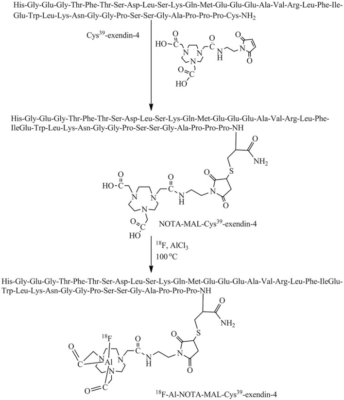
[68Ga]Ga-NOTA-MAL-Cys39-exendin-4, a potential GLP-1R targeted PET tracer for the detection of insulinoma.Nucl Med Biol. 2019 Jul-Aug;74-75:19-24. doi: 10.1016/j.nucmedbio.2019.08.002. Epub 2019 Aug 16. Nucl Med Biol. 2019. PMID: 31450071
-
Biased agonism and polymorphic variation at the GLP-1 receptor: Implications for the development of personalised therapeutics.Pharmacol Res. 2022 Oct;184:106411. doi: 10.1016/j.phrs.2022.106411. Epub 2022 Aug 22. Pharmacol Res. 2022. PMID: 36007775 Review.
Cited by
-
Evaluating the effectiveness and underlying mechanisms of incretin-based treatments for hypothalamic obesity: A narrative review.Obes Pillars. 2024 Feb 24;10:100104. doi: 10.1016/j.obpill.2024.100104. eCollection 2024 Jun. Obes Pillars. 2024. PMID: 38463533 Free PMC article. Review.
References
-
- Bertilsson G., Patrone C., Zachrisson O., Andersson A., Dannaeus K., Heidrich J., et al. (2008). Peptide hormone exendin-4 stimulates subventricular zone neurogenesis in the adult rodent brain and induces recovery in an animal model of Parkinson's disease. J. Neurosci. Res. 86 (2), 326–338. 10.1002/jnr.21483 - DOI - PubMed
Publication types
LinkOut - more resources
Full Text Sources