

Facial fillers: Relevant anatomy, injection techniques, and complications
- PMID: 37780674
- PMCID: PMC10541169
- DOI: 10.1002/wjo2.126
Facial fillers: Relevant anatomy, injection techniques, and complications
Abstract
Objective: The aim of this review article is to discuss the currently available facial fillers, their differences and indications, relevant anatomy, injection techniques, and avoidance and management of complications.
Data sources: Clinical experience and scientific papers.
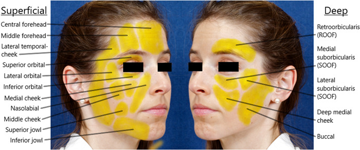
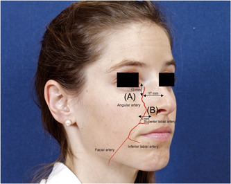
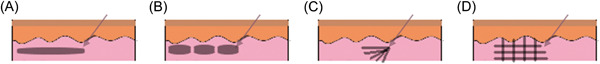
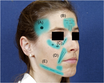
Conclusions: Reversal of facial aging via filler injection has been around since the late 1800s with the initial use of detrimental products. Today, many safe and effective products exist and can be tailored to the individual patient's desired effect. With the evolution of both products and injection techniques, the rate of complications with facial filler use is low. Nonetheless, providers offering facial filler injections should have detailed knowledge of facial anatomy, including facial planes and soft tissue compartments. Multiple injection techniques exist. Different techniques should be used, depending on the anatomic target. Providers should also know how to avoid and manage complications.
Keywords: cosmetic; facial; filler; injection; plastic surgery.
© 2023 The Authors. World Journal of Otorhinolaryngology ‐ Head and Neck Surgery published by John Wiley & Sons Ltd on behalf of Chinese Medical Association.
Conflict of interest statement
The authors declare no conflict of interest.
Figures
References
-
- Plastic Surgery Statistics Report 2020 . American Society of Plastic Surgeons National Clearinghouse of Plastic Surgery Procedural Statistics. 2020. Accessed October 20, 2022. https://www.plasticsurgery.org/documents/News/Statistics/2020/plastic-su...
-
- Surek CC. Facial anatomy for filler injection. Clin Plast Surg. 2019;46:603‐612. - PubMed
-
- Rohrich RJ, Pessa JE. The fat compartments of the face: anatomy and clinical implications for cosmetic surgery. Plast Reconstr Surg. 2007;119:2219‐2227. - PubMed
-
- Lee HJ, Won SY, O J, et al. The facial artery: a comprehensive anatomical review. Clin Anat (New York, N.Y.). 2018;31:99‐108. - PubMed
-
- Kontis T, Rivkin A. The history of injectable facial fillers. Facial Plast Surg. 2009;25:67‐72. - PubMed
Publication types
LinkOut - more resources
Full Text Sources