Review
. 2023 Sep 14:14:1250808.
doi: 10.3389/fneur.2023.1250808. eCollection 2023.

Imaging diagnosis in peripheral nerve injury

Affiliations
Review

Imaging diagnosis in peripheral nerve injury

Yanzhao Dong et al. Front Neurol. .

Abstract

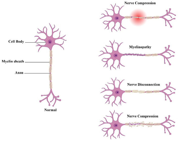
Peripheral nerve injuries (PNIs) can be caused by various factors, ranging from penetrating injury to compression, stretch and ischemia, and can result in a range of clinical manifestations. Therapeutic interventions can vary depending on the severity, site, and cause of the injury. Imaging plays a crucial role in the precise orientation and planning of surgical interventions, as well as in monitoring the progression of the injury and evaluating treatment outcomes. PNIs can be categorized based on severity into neurapraxia, axonotmesis, and neurotmesis. While PNIs are more common in upper limbs, the localization of the injured site can be challenging. Currently, a variety of imaging modalities including ultrasound (US), computed tomography (CT) and magnetic resonance imaging (MRI) and positron emission tomography (PET) have been applied in detection and diagnosis of PNIs, and the imaging efficiency and accuracy many vary based on the nature of injuries and severity. This article provides an overview of the causes, severity, and clinical manifestations of PNIs and highlights the role of imaging in their management.

Keywords: imaging; magnetic resonance imaging; peripheral nerve injury; positron emission tomography; ultrasound.


Conflict of interest statement

The authors declare that the research was conducted in the absence of any commercial or financial relationships that could be construed as a potential conflict of interest.

Figures

Figure 1
Figure 1
Neuropathic pain caused by different types of peripheral nerve damage.
Figure 2
Figure 2
US, MRI, pathological examination and intraoperative images of a 54-year-old female patient suffering from numbness and tenderness on the ulnar side of left hand. (A) US imaging on the left elbow showed hypoechoic thickening of the nerve fascicles at the cubital tunnel; (B) MRI showed significant thickening of the left ulnar nerve at the cubital tunnel with increased T2-weighted signal intensity. Red arrow marks the ulnar nerve swelling. O (olecranon), ME (medial epicondyle). (C) Pathological examination indicated small piece of fibrofatty tissue; (D) intraoperative image showed adhesion and edema of the ulnar nerve and surrounding tissue.
Figure 3
Figure 3
A case of patient with radial nerve entrapment. (A) US imaging of the radial nerve. Green star marks the proximal site of segmental entrapment. (B) Functional MRI of the radial nerve. Red triangle marks the site of radial nerve swelling. (C) Intraoperative image of deep branch of radial nerve. Segmental entrapment is observed. (D) MRI image of entrapped radial nerve. Yellow arrow makes the radial nerve swelling.
Figure 4
Figure 4
A case of patient suffering initially from allodynia and later a mass on the right elbow, which was pathologically confirmed as epithelioid sarcoma with ulnar nerve injury. (A) PET/CT showing the affected area; (B) coronal plane of the mass adjacent to ulnar nerve, red arrow marks the mass; (C) sagittal plane of the mass and the ulnar nerve, yellow arrow marks the swelling of the ulnar nerve; (D) intraoperative image of mass excision and nerve preservation.

References

    1. Hatzenbuehler J. Peripheral nerve injury. Curr Sports Med Rep. (2015) 14:356–7. doi: 10.1249/JSR.0000000000000186 - DOI - PubMed
    1. Menorca RM, Fussell TS, Elfar JC. Nerve physiology: mechanisms of injury and recovery. Hand Clin. (2013) 29:317–30. doi: 10.1016/j.hcl.2013.04.002, PMID: - DOI - PMC - PubMed
    1. Kumar A, Shukla D, Bhat DI, Devi BI. Iatrogenic peripheral nerve injuries. Neurol India. (2019) 67:S135–9. doi: 10.4103/0028-3886.250700, PMID: - DOI - PubMed
    1. Vijayavenkataraman S. Nerve guide conduits for peripheral nerve injury repair: a review on design, materials and fabrication methods. Acta Biomater. (2020) 106:54–69. doi: 10.1016/j.actbio.2020.02.003, PMID: - DOI - PubMed
    1. Yang H, Dong Y, Wang Z, Lai J, Yao C, Zhou H, et al. Traumatic neuromas of peripheral nerves: diagnosis, management and future perspectives. Front Neurol. (2022) 13:1039529. doi: 10.3389/fneur.2022.1039529 - DOI - PMC - PubMed

LinkOut - more resources