

Stillbirth Investigations: An Iconographic and Concise Diagnostic Workup in Perinatal Pathology
- PMID: 37780873
- PMCID: PMC10539070
- DOI: 10.1055/s-0043-1764485
Stillbirth Investigations: An Iconographic and Concise Diagnostic Workup in Perinatal Pathology
Abstract
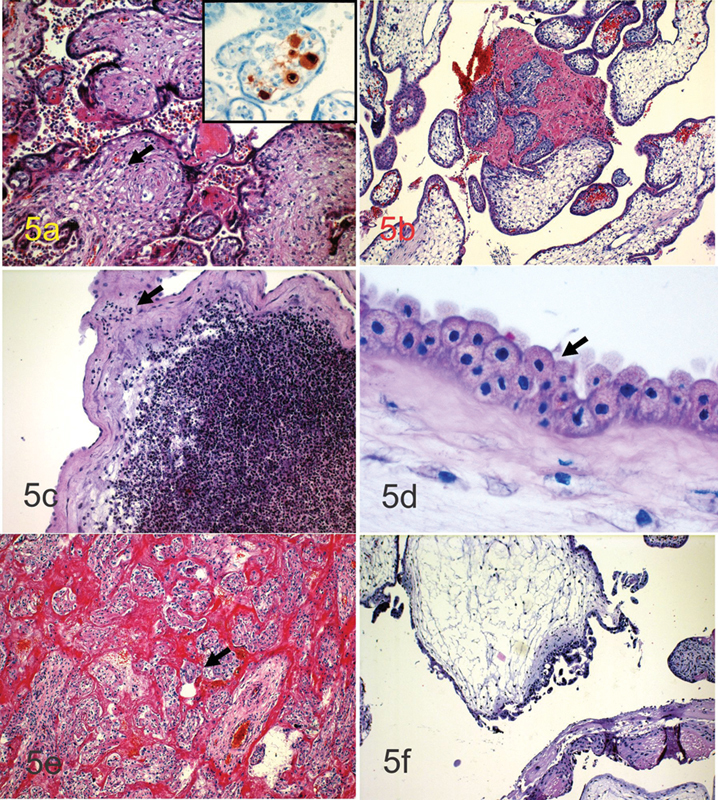
Introduction Stillbirth is a dramatic event for the parents, health care team, and anyone close to the expectant parents. Multidisciplinary team (MDT) meetings are essential to improve communication in health care. We review the most frequent findings discussed at MDT meetings. Methods A PubMed search was conducted through December 2021 since the inception (1965) using clinical queries with the key terms "stillbirth" AND "investigation" AND "pathology" AND "human." The search strategy included reviews, meta-analyses, randomized controlled trials, clinical trials, and observational studies. This systematic review is based on, but not limited to, the search results. It is the experience of more than 30 years of pediatrics, obstetrics, and pathology staff. Results Two hundred and six articles were screened and complemented through the perusal of congressional activities and personal communications. Pathological findings following perinatal death can be divided into macroscopic, histologic, and placental findings. The placenta is crucial in fetal medicine and is key in determining the cause of stillbirth in a substantial number of events. Perinatal lung disease is essential to evaluate the response of newborns to extrauterine life and address newborns' outcomes appropriately. Conclusions Stillbirth remains one of the less explored areas of medicine, and we can determine the cause in a limited number of cases. Nevertheless, placental pathology is critical in the etiology discovery pathway. Accurate investigations and discussion of photography-supported findings are vital in promoting communication at MDT meetings.
Keywords: Perinatal Investigation; early neonatal illness; intrapartum distress; stillbirth.
The Indian Association of Laboratory Physicians. This is an open access article published by Thieme under the terms of the Creative Commons Attribution-NonDerivative-NonCommercial License, permitting copying and reproduction so long as the original work is given appropriate credit. Contents may not be used for commercial purposes, or adapted, remixed, transformed or built upon. ( https://creativecommons.org/licenses/by-nc-nd/4.0/ ).
Conflict of interest statement
Conflict of Interest None declared.
Figures
Similar articles
-
The future of Cochrane Neonatal.Early Hum Dev. 2020 Nov;150:105191. doi: 10.1016/j.earlhumdev.2020.105191. Epub 2020 Sep 12. Early Hum Dev. 2020. PMID: 33036834
-
Care prior to and during subsequent pregnancies following stillbirth for improving outcomes.Cochrane Database Syst Rev. 2018 Dec 17;12(12):CD012203. doi: 10.1002/14651858.CD012203.pub2. Cochrane Database Syst Rev. 2018. PMID: 30556599 Free PMC article.
-
Living with Loss: study protocol for a randomized controlled trial evaluating an internet-based perinatal bereavement program for parents following stillbirth and neonatal death.Trials. 2022 Jun 6;23(1):464. doi: 10.1186/s13063-022-06363-0. Trials. 2022. PMID: 35668502 Free PMC article.
-
Diagnostic utility of serial circulating placental growth factor levels and uterine artery Doppler waveforms in diagnosing underlying placental diseases in pregnancies at high risk of placental dysfunction.Am J Obstet Gynecol. 2022 Oct;227(4):618.e1-618.e16. doi: 10.1016/j.ajog.2022.05.043. Epub 2022 May 27. Am J Obstet Gynecol. 2022. PMID: 35644246
-
Parameters for estimating the time of death at perinatal autopsy of stillborn fetuses: a systematic review.Int J Legal Med. 2019 Mar;133(2):483-489. doi: 10.1007/s00414-019-01999-1. Epub 2019 Jan 8. Int J Legal Med. 2019. PMID: 30617766
Cited by
-
Fetal Death Due to an Unusual Coexistence of Two Umbilical Cord Anomalies: Analysis in a Forensic Perspective.Diagnostics (Basel). 2025 Jun 3;15(11):1423. doi: 10.3390/diagnostics15111423. Diagnostics (Basel). 2025. PMID: 40506995 Free PMC article.
References
-
- Corabian P, Scott N A, Lane C, Guyon G. Guidelines for investigating stillbirths: an update of a systematic review. J Obstet Gynaecol Can. 2007;29(07):560–567. - PubMed
-
- Pinar H. Postmortem findings in term neonates. Semin Neonatol. 2004;9(04):289–302. - PubMed
-
- Sergi C M, Mullur T. Life and death sometimes coincide, and pastoral response is crucial to the brokenhearted. J Pastoral Care Counsel. 2022;76(04):281–284. - PubMed
Publication types
LinkOut - more resources
Full Text Sources