

Thrombus or tumor?
- PMID: 37780915
- PMCID: PMC10539678
- DOI: 10.1002/ccr3.7975
Thrombus or tumor?
Abstract
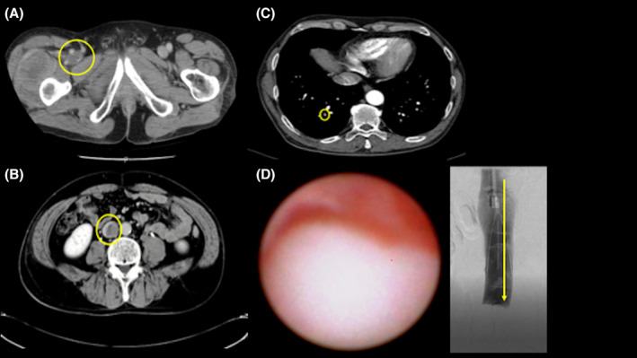
Key clinical message: Contrast defects in veins are often diagnosed as benign thrombi, but depending on the patient's background it is necessary to differentiate between tumor thrombi. It is difficult to differentiate between these using contrast-enhanced CT alone, but with angioscopy it is easy to visually distinguish between a benign and tumor thrombi.
Abstract: Contrast-enhanced computer tomography (CT) performed on a male patient being treated for de-differentiated chondrosarcoma revealed contrast defects in the pulmonary artery and right femoral vein, and a diagnosis of pulmonary artery thromboembolism and venous thromboembolism was made, and oral anticoagulant therapy was started. However, a follow-up CT showed that the contrast defect had extended to the inferior vena cava. Observation using an angioscope revealed that it was not a benign thrombi but a tumor.
Keywords: angioscopy; deep vein thrombosis; pulmonary embolism; tumor thrombus.
© 2023 The Authors. Clinical Case Reports published by John Wiley & Sons Ltd.
Conflict of interest statement
The authors declare that they have no conflict of interests.
Figures
References
-
- Sharma P, Kumar R, Jeph S, et al. 18F‐FDG PET‐CT in the diagnosis of tumor thrombus: can it be differentiated from benign thrombus? Nucl Med Commun. 2011;32(9):782‐788. - PubMed
-
- Uchida Y, Nakamura F, Tsukamoto M, You S, Kido H, Sugimoto T. Percutaneous ventricular endomyocardial biopsy with angioscopic guidance. Am Heart J. 1989;118(5 Pt 1):1039‐1041. - PubMed
Publication types
LinkOut - more resources
Full Text Sources
