

Controversies and progress on standardization of large-scale brain network nomenclature
- PMID: 37781138
- PMCID: PMC10473266
- DOI: 10.1162/netn_a_00323
Controversies and progress on standardization of large-scale brain network nomenclature
Abstract
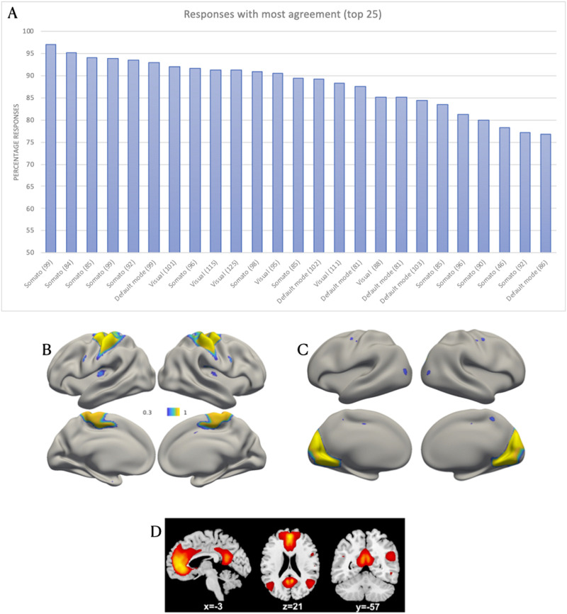
Progress in scientific disciplines is accompanied by standardization of terminology. Network neuroscience, at the level of macroscale organization of the brain, is beginning to confront the challenges associated with developing a taxonomy of its fundamental explanatory constructs. The Workgroup for HArmonized Taxonomy of NETworks (WHATNET) was formed in 2020 as an Organization for Human Brain Mapping (OHBM)-endorsed best practices committee to provide recommendations on points of consensus, identify open questions, and highlight areas of ongoing debate in the service of moving the field toward standardized reporting of network neuroscience results. The committee conducted a survey to catalog current practices in large-scale brain network nomenclature. A few well-known network names (e.g., default mode network) dominated responses to the survey, and a number of illuminating points of disagreement emerged. We summarize survey results and provide initial considerations and recommendations from the workgroup. This perspective piece includes a selective review of challenges to this enterprise, including (1) network scale, resolution, and hierarchies; (2) interindividual variability of networks; (3) dynamics and nonstationarity of networks; (4) consideration of network affiliations of subcortical structures; and (5) consideration of multimodal information. We close with minimal reporting guidelines for the cognitive and network neuroscience communities to adopt.
Keywords: Brain network; Cognitive neuroscience; Diffusion weighted imaging; EEG; Functional connectivity; MEG; Network neuroscience; Parcellation; Resting-state fMRI; Structural connectivity.
© 2023 Massachusetts Institute of Technology.
Figures

Similar articles
-
Mean-Field Modeling of Brain-Scale Dynamics for the Evaluation of EEG Source-Space Networks.Brain Topogr. 2022 Jan;35(1):54-65. doi: 10.1007/s10548-021-00859-9. Epub 2021 Jul 9. Brain Topogr. 2022. PMID: 34244910
-
Brain parcellation driven by dynamic functional connectivity better capture intrinsic network dynamics.Hum Brain Mapp. 2021 Apr 1;42(5):1416-1433. doi: 10.1002/hbm.25303. Epub 2020 Dec 7. Hum Brain Mapp. 2021. PMID: 33283954 Free PMC article.
-
Brain parcellation selection: An overlooked decision point with meaningful effects on individual differences in resting-state functional connectivity.Neuroimage. 2021 Nov;243:118487. doi: 10.1016/j.neuroimage.2021.118487. Epub 2021 Aug 19. Neuroimage. 2021. PMID: 34419594 Free PMC article.
-
Issues and recommendations from the OHBM COBIDAS MEEG committee for reproducible EEG and MEG research.Nat Neurosci. 2020 Dec;23(12):1473-1483. doi: 10.1038/s41593-020-00709-0. Epub 2020 Sep 21. Nat Neurosci. 2020. PMID: 32958924 Review.
-
Large-scale functional connectivity networks in the rodent brain.Neuroimage. 2016 Feb 15;127:496-509. doi: 10.1016/j.neuroimage.2015.12.017. Epub 2015 Dec 17. Neuroimage. 2016. PMID: 26706448 Review.
Cited by
-
Default Mode Network Functional Connectivity As a Transdiagnostic Biomarker of Cognitive Function.Biol Psychiatry Cogn Neurosci Neuroimaging. 2025 Apr;10(4):359-368. doi: 10.1016/j.bpsc.2024.12.016. Epub 2025 Jan 9. Biol Psychiatry Cogn Neurosci Neuroimaging. 2025. PMID: 39798799 Review.
-
Reduced lateralization of multiple functional brain networks in autistic males.J Neurodev Disord. 2024 May 8;16(1):23. doi: 10.1186/s11689-024-09529-w. J Neurodev Disord. 2024. PMID: 38720286 Free PMC article.
-
Domain-Specific Prediction of Clinical Progression in Parkinson's Disease Using the Mosaic Approach.Brain Behav. 2025 Jan;15(1):e70289. doi: 10.1002/brb3.70289. Brain Behav. 2025. PMID: 39789902 Free PMC article.
-
Functional differentiation of the default and frontoparietal control networks predicts individual differences in creative achievement: evidence from macroscale cortical gradients.Cereb Cortex. 2025 Mar 6;35(3):bhaf046. doi: 10.1093/cercor/bhaf046. Cereb Cortex. 2025. PMID: 40056422
-
Effects of Oral Contraceptive Pills on Brain Networks: A Conceptual Replication and Extension.Hum Brain Mapp. 2025 Aug 1;46(11):e70318. doi: 10.1002/hbm.70318. Hum Brain Mapp. 2025. PMID: 40782033 Free PMC article. Clinical Trial.
References
-
- Anderson, K. M., Ge, T., Kong, R., Patrick, L. M., Spreng, R. N., Sabuncu, M. R., Yeo, B. T. T., & Holmes, A. J. (2021). Heritability of individualized cortical network topography. Proceedings of the National Academy of Sciences of the United States of America, 118(9), e2016271118. 10.1073/pnas.2016271118, - DOI - PMC - PubMed
Grants and funding
- R01 MH112517/MH/NIMH NIH HHS/United States
- P50 HD103573/HD/NICHD NIH HHS/United States
- R01 EB021265/EB/NIBIB NIH HHS/United States
- R01 MH116226/MH/NIMH NIH HHS/United States
- R01 MH071589/MH/NIMH NIH HHS/United States
- R01 MH118370/MH/NIMH NIH HHS/United States
- R01 AG068563/AG/NIA NIH HHS/United States
- U01 EB026996/EB/NIBIB NIH HHS/United States
- R01 NS119911/NS/NINDS NIH HHS/United States
- P50 MH106435/MH/NIMH NIH HHS/United States
- R01 MH119091/MH/NIMH NIH HHS/United States
- R21 NS104603/NS/NINDS NIH HHS/United States
- U01 DA050987/DA/NIDA NIH HHS/United States
LinkOut - more resources
Full Text Sources