. 2023 Sep 13:14:1228488.
doi: 10.3389/fphys.2023.1228488. eCollection 2023.

p90RSK2, a new MLCK mediates contractility in myosin light chain kinase null smooth muscle

Affiliations

p90RSK2, a new MLCK mediates contractility in myosin light chain kinase null smooth muscle

Jaspreet Kalra et al. Front Physiol. .

Abstract

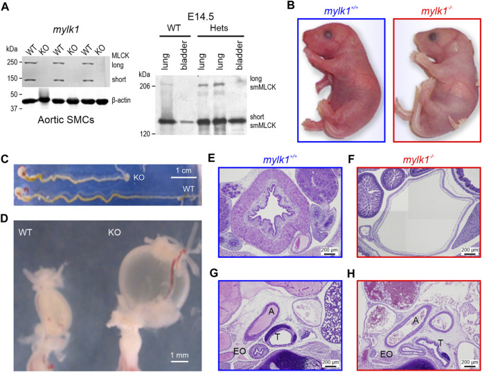
Introduction: Phosphorylation of smooth muscle (SM) myosin regulatory light chain (RLC20) is a critical switch leading to SM contraction. The canonical view held that only the short isoform of myosin light chain kinase (MLCK1) catalyzed this reaction. It is now accepted that auxiliary kinases may contribute to vascular SM tone and contractility. We have previously reported that p90 ribosomal S6 kinase (RSK2) functions as such a kinase, in parallel with MLCK1, contributing ∼25% of the maximal myogenic force in resistance arteries. Thus, RSK2 may be instrumental in the regulation of basal vascular tone and blood pressure. Here, we take advantage of a MLCK1 null mouse (mylk1 -/-) to further test our hypothesis that RSK2 can function as an MLCK, playing a significant physiological role in SM contractility. Methods: Using fetal (E14.5-18.5) SM tissues, as embryos die at birth, we investigated the necessity of MLCK for contractility and fetal development and determined the ability of RSK2 kinase to compensate for the lack of MLCK and characterized its signaling pathway in SM. Results and Discussion: Agonists induced contraction and RLC20 phosphorylation in mylk1 -/- SM was attenuated by RSK2 inhibition. The pCa-tension relationships in permeabilized strips of bladder showed no difference in Ca2+ sensitivity in WT vs mylk1 -/- muscles, although the magnitude of force responses was considerably smaller in the absence of MLCK. The magnitude of contractile responses was similar upon addition of GTPγS to activate the RhoA/ROCK pathway or calyculinA to inhibit the myosin phosphatase. The Ca2+-dependent tyrosine kinase, Pyk2, contributed to RSK2-mediated contractility and RLC20 phosphorylation. Proximity-ligation and immunoprecipitation assays demonstrated an association of RSK2, PDK1 and ERK1/2 with MLCK and actin. RSK2, PDK1, ERK1/2 and MLCK formed a signaling complex on the actin filament, positioning them for interaction with adjacent myosin heads. The Ca2+-dependent component reflected the agonist mediated increases in Ca2+, which activated the Pyk2/PDK1/RSK2 signaling cascade. The Ca2+-independent component was through activation of Erk1/2/PDK1/RSK2 leading to direct phosphorylation of RLC20, to increase contraction. Overall, RSK2 signaling constitutes a new third signaling pathway, in addition to the established Ca2+/CaM/MLCK and RhoA/ROCK pathways to regulate SM contractility.

Keywords: MLCK; PDK1; Pyk2; RSK2; mylk1; p90 ribosomal S6 kinase; smooth muscle.


Conflict of interest statement

Author AF was employed by Brain Surgery Worldwide. The remaining authors declare that the research was conducted in the absence of any commercial or financial relationships that could be construed as a potential conflict of interest.

Figures

FIGURE 1
FIGURE 1
Long (220 kDa) and short (130 kDa) MLCK and telokin (17 kDa) protein expression and phenotypic differences in mylk1 −/− and WT littermate mice. (A) Left panel: Western blots showing complete loss of MLCK isoforms 130 and 220 kDa in aortic SM cells cultured from E18.5 mylk1 −/− mice. Loss of 17 kDa telokin is shown in Supplementary Figure S1B. Right panel: 220 kDa MLCK was not detected in E14.5 WT or mylk +/− bladder, with lung serving as a positive control. Proteins were subjected to SDS-PAGE followed by Western blotting with the anti–smMLCK (1:10000) antibodies and fluorescent Alexa Fluor 680 anti–mouse IgG secondary antibody (1:15000) and visualized by LICOR Imager. (B) Postnatal pups. Mylk1 −/− died shortly after birth. Mylk1 −/− pups are typically paler, the length of the gut, stomach to rectum is shorter (C) and the bladder is markedly enlarged (D–F). Histology shows other dilated smooth muscle containing organs in transverse sections of E18.5 day embryos, stained with hematoxylin and eosin (G,H). A, aorta; T, trachea; EO, esophagus. The image in (F) is composed of four images stitched together in order to match the magnification of the panels (E,G,H).
FIGURE 2
FIGURE 2
Assays demonstrating that Mylk −/− SM retained contractile activity: (A) Umbilical arteries from WT and mylk1 −/− E18.5 embryos developed different magnitudes of force in response to high [K+]. Following permeabilization with α-toxin, phosphatase inhibitor calyculin A (100 nM) induced comparable magnitudes of maximal force in the mylk1 −/− and the WT littermate vessels in pCa > 8 EGTA buffered solution. Ratios of calyculin A high [K+] in the graph demonstrate the reduced high [K+] compared to maximal force in the mylk1 −/− compared to WT. (B) Different rates of contraction in response to high [K+] occur in WT compared to mylk1 −/− in both bladder and umbilical arteries. Graph, the half-time of high [K+] contractions in mylk1 −/− SM strips was significantly greater than wildtype in both bladder (p < 0.0001) and arteries (p < 0.05). (C) Mylk1 −/− SM contracts in responses to increasing [Ca2+]i, in α-toxin permeabilized bladder E18.5 mylk1 −/− vs. WT SM strips. Left panel, pCa-tension responses. Graph, pCa-tension curve normalized to force at pCa 4.5 (100%). The magnitude of contractile force was less but the sensitivity to Ca2+ was not significantly different in mylk1 −/− vs. WT bladder SM (n = 10 and 3 biological replicates respectively). (D) Activation of the RhoA/ROCK signaling pathway by addition of 10 μM GTPγS to α-toxin permeabilized WT and mylk1 −/− bladder in pCa 6.3 solution. The ROCK inhibitor, Y27632, 5 and 10 μM relaxed the GTPγS-induced contractions. Graphs showed that the magnitude of GTPγS-induced contractions did not significantly differ between mylk1 −/− and WT SM (n = 4 and 5 biological replicates respectively) but that the Y27632-induced relaxation of GTPγS-induced contractions was greater in the mylk1 −/− SM compared to WT (p < 0.01, n = 5 and 6 biological replicates respectively).
FIGURE 3
FIGURE 3
RSK2 kinase mediates myosin RLC20 phosphorylation in mylk1 −/− SM cells. (A) LPA stimulation of cultured embryonic aortic mylk1 −/− and mylk1 +/+ cells resulted in an increase in RLC20 phosphorylation (n = 3 biological replicates). (B) RSK inhibitor, LJH685, significantly inhibited RLC20 phosphorylation in mylk1 −/− SM cells stimulated with LPA (p < 0.01, n = 4 biological replicates) or with thromboxane analogue, U47619 (p < 0.001, n = 3 biological replicates).
FIGURE 4
FIGURE 4
RSK inhibitor BiD1870 inhibited contractile responses to carbachol in mylk1 −/− and WT bladder SM. (A) Summary of magnitude of high [K+]-induced phasic and tonic components of contraction in WT (n = 6) and mylk1 −/− (n = 4) bladder SM. The high [K+] phasic component was absent or small in the mylk1 −/− while the tonic components present in embryonic bladder did not differ significantly in magnitude p = 0.5. (B) The contractile response to high [K+] was recorded and following return to high [Na+] Hepes buffered Krebs solution the muscles were treated with increasing concentrations of carbachol (Cch) in the presence or absence of the RSK inhibitor, BiD 1870 (1 μM) or equivalent volumes of diluent, DMSO. (C) Contractile responses to increasing concentrations of Cch were normalized to the tonic high [K+] contractions for each muscle and analyzed by 2-way Anova. WT DMSO vs. WT BiD 1870: p < 0.0001, mylk1 −/− DMSO vs. mylk1 −/− BiD 1870: p < 0.003, n = 3 WT, n = 2 mylk1 −/−. Arrows = incremental Cch concentrations from 5 × 10⁻⁹ to 5 × 10⁻⁵.
FIGURE 5
FIGURE 5
RSK2 and upstream activators, ERK1/2 and PDK1 associate with MLCK and actin in SM cells: (A) RSK2 and MLCK colocalized in the proximity ligation assay (PLA) in WT, but not RSK2−/− and mylk1 −/− SM cells, in both serum starved or in the presence of serum plus the thromboxane analogue, U46619. Cells were labeled with rabbit monoclonal anti-MLCK antibody (1:200) and mouse monoclonal anti-RSK2 antibody (1:200), followed by secondary antibodies coupled to oligonucleotides (PLA probes). Following amplification of PLA probes in close proximity, complementary oligos coupled to fluorochromes hybridized to the amplicons and gave rise to discrete fluorescent puncta. Blue nuclei are stained with DAPI. The number of puncta/cell was significantly greater in the serum plus U46619 stimulated WT cells compared to non-stimulated (p < 0.001 n = 3 biological replicates) and to a lesser extent in non-stimulated WT cells compared to non-stimulated mylk1 −/− SM cells (<0.05 n = 3 biological replicates). RSK−/− and mylk1 −/− cells served as negative controls and had few puncta with or without stimulation. (B) MLCK immunoprecipitation (IP). RSK2 and actin were immunoprecipitated by MLCK in WT but not RSK2−/− SM cells or beads alone. Neither RSK2, MLCK or actin were immunoprecipitated by IgG alone (Supplementary Figure S1C). (C) Representative Western blot and graph showing MLCK IP from mouse WT aortic SM cells in the absence or presence of serum. RSK2, phosphorylated RSK2S227, phosphorylated ERK1/2 and phosphorylated PDK1Ser241 as well as actin were immunoprecipitated by MLCK in both the presence and absence of serum (n = 4 biological replicates). Serum conditions significantly increased the amount of phosphorylated RSK2S227 immunoprecipitated by MLCK p < 0.001, while there was an increased trend with phosphorylated ERK1/2. Phosphorylated proteins were normalized to actin and 0 serum conditions taken as 1 for each phosphorylated protein in the graph.
FIGURE 6
FIGURE 6
AngII stimulation increased phosphorylation of the Ca2+ dependent tyrosine kinase, Pyk2 and of its downstream targets PDK1 and RSK2 which was suppressed by Pyk2 inhibitor, PF-4618433. (A) Representative Western blot and graphs from cultured E18.5 aortic SM cells showing significant AngII-induced phosphorylation of both Pyk2 (n = 8 biological replicates, p < 0.001) and PDK1 (n = 7 biological replicates, p < 0.01). The Pyk2 inhibitor, (PF-4618433), inhibited activation of both Pyk2 and its downstream target, PDK1 (n = 8, p < 0.001 and n = 7, p < 0.01 respectively). (B) AngII increased RSK2 phosphorylation (n = 5, p < 0.05) that was significantly inhibited by PF-4618433 (n = 5, p < 0.01). Proteins normalized to actin. (C) Three signaling pathways regulating SM RLC20 phosphorylation, contractility, basal tone and regulation of blood pressure. Agonists, neurotransmitters or increases in arterial intraluminal pressure activate; 1) the canonical Ca2+ activated MLCK pathway. 2) the RhoA/ROCK pathway that phosphorylates and inhibits MLCP resulting in an increase in RLC20 phosphorylation. 3) the new RSK2 pathway that leads to activation of NHE-1 resulting in cytosolic alkalinization and increase in Ca2+ events. This increased Ca2+ in addition to Ca2+influx and release from stores, feeds back to further activated the MLCK pathway. Activated RSK2 can also directly phosphorylate RLC20. Different stimuli may preferentially select for a given signaling pathway. Created by BioRender.com.

Update of

References

    1. Amano M., Ito M., Kimura K., Fukata Y., Chihara K., Nakano T., et al. (1996). Phosphorylation and activation of myosin by Rho-associated kinase (Rho-kinase). J. Biol. Chem. 271, 20246–20249. 10.1074/jbc.271.34.20246 - DOI - PubMed
    1. Anjum R., Blenis J. (2008). The RSK family of kinases: emerging roles in cellular signalling. Nat. Rev. Mol. Cell Biol. 9, 747–758. nrm2509 [pii]. 10.1038/nrm2509 - DOI - PubMed
    1. Artamonov M., Momotani K., Utepbergenov D., Franke A., Khromov A., Derewenda Z. S., et al. (2013a). The p90 ribosomal S6 kinase (RSK) is a mediator of smooth muscle contractility. PLoS One 8, e58703. PONE-D-12-30703 [pii]. 10.1371/journal.pone.0058703 - DOI - PMC - PubMed
    1. Artamonov M. V., Momotani K., Stevenson A., Trentham D. R., Derewenda U., Derewenda Z. S., et al. (2013b). Agonist-induced Ca2+ sensitization in smooth muscle: redundancy of rho guanine nucleotide exchange factors (RhoGEFs) and response kinetics, a caged compound study. J. Biol. Chem. 288, 34030–34040. [pii]. 10.1074/jbc.M113.514596 - DOI - PMC - PubMed
    1. Artamonov M. V., Sonkusare S. K., Good M. E., Momotani K., Eto M., Isakson B. E., et al. (2018). RSK2 contributes to myogenic vasoconstriction of resistance arteries by activating smooth muscle myosin and the Na(+)/H(+) exchanger. Sci. Signal 11, eaar3924. 10.1126/scisignal.aar3924 - DOI - PMC - PubMed