. 2023 Sep 14:10:1208919.
doi: 10.3389/fvets.2023.1208919. eCollection 2023.

Lymphocytic hypophysitis in dogs infected with Leishmania spp

Affiliations

Lymphocytic hypophysitis in dogs infected with Leishmania spp

Edenilson Doná Frigerio et al. Front Vet Sci. .

Abstract

Background: Morphological involvement of endocrine glands, such as the pituitary gland, remain uninvestigated in dogs with canine visceral leishmaniasis. Therefore, this study investigated the presence of amastigotes of Leishmania spp. and characterized inflammatory changes, highlighting the involvement of TCD3+ lymphocytes in different regions of the pituitary gland of dogs.

Methods: Samples were collected from 21 naturally infected dogs and 5 control, uninfected dogs. The different pituitary regions were analyzed in histological sections stained with hematoxylin and eosin (HE) under light microscopy. Inflammation was classified by intensity in a score from 0 to 3, absent (0), mild (1), moderate (2), and marked (3). The immunohistochemical (IHC) evaluation was performed in five high-power fields (hot spot) in a 40x objective of each region with manual counting (Image J1.52ª) of the TCD3+ lymphocytes and for amastigotes analyzed in 40x and 100x objectives. The Shapiro-Wilk test was used to assess the normality of the data. Differences between groups were determined by the Mann Whitney test. The correlation between variables was assessed by Sperman's correlation test. p < 0.05 were considered statistically significant.

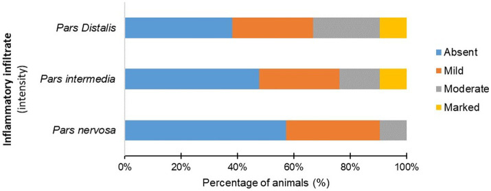
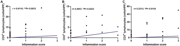
Results: Amastigotes from the pituitary glands of two infected dogs were identified using IHC. The histopathological evaluation stained with hematoxylin and eosin showed greater intensity of inflammation in the pars distalis and pars intermedia regions of infected dogs. IHC for TCD3+ lymphocytes showed a higher median number of immunolabeled cells in pars nervosa in the infected group than in the control group (p < 0.05); and expecting a variation in the distribution and number of these cells in naturally infected dogs, the median of the control group was considered a cut-off point, an increase in T lymphocytes (p < 0.05) was also observed in the pars intermedia and pars distalis of an infected subgroup (n = 10). A moderate significant correlation between the intensity of inflammation and the number of immunolabeled TCD3+ lymphocytes was established in the analyzed pituitary regions, characterizing the occurrence of hypophysitis.

Conclusion: These findings presuppose that inflammation and/or the parasite in the pituitary region can result in gland dysfunction, worsening the clinical condition of the patient and compromising the efficiency of treatment and prognosis.

Keywords: Leishmania infantum; T lymphocyte; immunohistochemical; inflammation; pituitary gland.


Conflict of interest statement

The authors declare that the research was conducted in the absence of any commercial or financial relationships that could be construed as a potential conflict of interest.

Figures

Figure 1
Figure 1
Photomicrography of the pituitary gland of a dog infected with Leishmania spp. Immunohistochemistry for detection of amastigotes (arrows). Positive immunolabeling amastigotes in the cytoplasm of macrophages and free in pars intermedia (PI) of the adenohypophysis in the marginal area with pars nervosa (PN; hyperimmune mouse serum +3,3′-diaminobezidine- DAKO; scale bar = 20 μm).
Figure 2
Figure 2
Photomicrograph of the pituitary regions of the dogs (G1), demonstrating the distribution and intensity of inflammation composed of mononuclear cells (arrows). (A) Note the inflammatory cells with focal, perivascular distribution in the pars nervosa (PN), with moderate intensity. (B) Observe inflammatory cells intermingled with chromophilic, acidophilic, and basophilic cells with focal distribution and intensity in pars distalis (PD), and absence of inflammation in pars intermedia (PI). (C) Note the inflammation with focal and moderate distribution in the parenchyma in pars distalis (HE staining, bar = 20 μm).
Figure 3
Figure 3
Percentage (%) of infected dogs (G1) according to the intensity of mononuclear inflammatory cells observed in the pars distalis, pars intermedia, and pars nervosa.
Figure 4
Figure 4
(A,C,E) Photomicrograph representing the pars nervosa (PN), pars intermedia (PI), and pars distalis regions of the pituitary gland of a dog infected with Leishmania spp., immunohistochemistry for detection of CD3+ T lymphocytes revealed with DAB DAKO (scale bar = 20 μm). (B,D,F) Dispersion graphs showing the number of TCD3+ lymphocytes in the pituitary regions of dogs in group G1 (n = 21) and G2 (control n = 5). The horizontal lines represent the median and interquartile range values. (B) The number of TCD3+ cells was higher in the pars nervosa (*p = 0.0357). (D,F) No difference in the pars intermedia (p = 0.4935) and pars distalis (p = 0.4000).
Figure 5
Figure 5
Dispersion graphs showing the number of TCD3+ lymphocytes in the pituitary regions of G1 subpopulations. The horizontal lines represent the median and interquartile range values. (A) In the pars nervosa evaluation, the animal with the highest number of TCD3+ cells was removed from the infected dogs (G1; n = 20), and when compared to the control group (G2; n = 5), the statistical difference was maintained (* p = 0.0441); (B,C) the pars intermedia and pars distalis evaluation resulted in a subpopulation of ten infected dogs (G1; n = 10) with higher numbers of TCD3+ cells compared to the control group (G2; n = 5) being selected. This subpopulation of dogs showed significantly increased TCD3+ cells, respectively (** p = 0.0070) and (*** p = 0.0007).
Figure 6
Figure 6
Dispersion graphs showing a moderate positive correlation between inflammation score and median TCD3+ lymphocyte count per pituitary region in the infected group (G1, n = 21) using Spearman’s test. (A) Pars nervosa (r = 0.6110; **p = 0.0033). (B) Pars intermedia (r = 0.4957; *p = 0.0223). (C) Pars distalis (r = 0.5213; *p = 0.0154).

References

    1. Perez-Castro C, Renner U, Haedo MR, Stalla GK, Arzt E. Cellular and molecular specificity of pituitary gland physiology. Physiol Rev. (2012) 92:1–38. doi: 10.1152/physrev.00003.2011 - DOI - PubMed
    1. Polledo L, Grinwis GCM, Graham P, Dunning M, Baiker K. Pathological findings in the pituitary glands of dogs and cats. Vet Pathol. (2018) 55:880–8. doi: 10.1177/0300985818784162 - DOI - PubMed
    1. Miller MA, Bruyette DS, Scott-Moncrieff JC, Owen TJ, Ramos-Vara JA, Weng HY, et al. Histopathologic findings in canine pituitary glands. Vet Pathol. (2018) 55:871–9. doi: 10.1177/0300985818766211, PMID: - DOI - PubMed
    1. Bellastella A, Bizzarro A, Coronella C, Bellastella G, Sinisi AA, De Bellis A. Lymphocytic hypophysitis: a rare or underestimated disease? Eur J Endocrinol. (2003) 149:363–76. doi: 10.1530/eje.0.1490363 - DOI - PubMed
    1. Langlois F, Varlamov EV, Fleseriu M. Hypophysitis, the growing spectrum of a rare pituitary disease. J Clin Endocrinol Metab. (2022) 107:10–28. doi: 10.1210/clinem/dgab672, PMID: - DOI - PMC - PubMed