

The functional response of human monocyte-derived macrophages to serum amyloid A and Mycobacterium tuberculosis infection
- PMID: 37781389
- PMCID: PMC10540855
- DOI: 10.3389/fimmu.2023.1238132
The functional response of human monocyte-derived macrophages to serum amyloid A and Mycobacterium tuberculosis infection
Abstract
Introduction: In the course of tuberculosis (TB), the level of major acute phase protein, namely serum amyloid A (hSAA-1), increases up to a hundredfold in the pleural fluids of infected individuals. Tubercle bacilli infecting the human host can be opsonized by hSAA-1, which affects bacterial entry into human macrophages and their intracellular multiplication.
Methods: We applied global RNA sequencing to evaluate the functional response of human monocyte-derived macrophages (MDMs), isolated from healthy blood donors, under elevated hSAA-1 conditions and during infection with nonopsonized and hSAA-1-opsonized Mycobacterium tuberculosis (Mtb). In the same infection model, we also examined the functional response of mycobacteria to the intracellular environment of macrophages in the presence and absence of hSAA-1. The RNASeq analysis was validated using qPCR. The functional response of MDMs to hSAA-1 and/or tubercle bacilli was also evaluated for selected cytokines at the protein level by applying the Milliplex system.
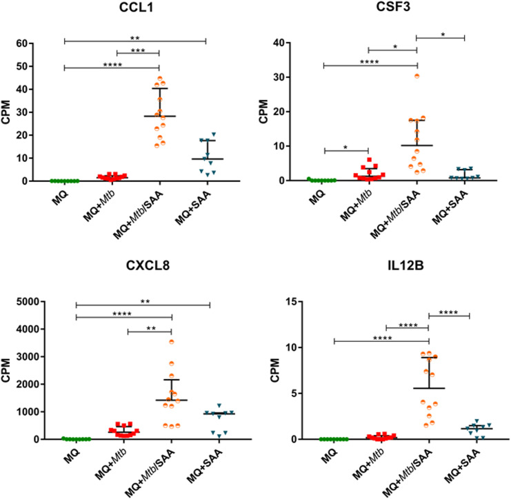
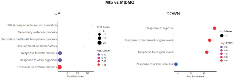
Findings: Transcriptomes of MDMs cultured in the presence of hSAA-1 or infected with Mtb showed a high degree of similarity for both upregulated and downregulated genes involved mainly in processes related to cell division and immune response, respectively. Among the most induced genes, across both hSAA-1 and Mtb infection conditions, CXCL8, CCL15, CCL5, IL-1β, and receptors for IL-7 and IL-2 were identified. We also observed the same pattern of upregulated pro-inflammatory cytokines (TNFα, IL-6, IL-12, IL-18, IL-23, and IL-1) and downregulated anti-inflammatory cytokines (IL-10, TGFβ, and antimicrobial peptide cathelicidin) in the hSAA-1 treated-MDMs or the phagocytes infected with tubercle bacilli. At this early stage of infection, Mtb genes affected by the inside microenvironment of MDMs are strictly involved in iron scavenging, adaptation to hypoxia, low pH, and increasing levels of CO2. The genes for the synthesis and transport of virulence lipids, but not cholesterol/fatty acid degradation, were also upregulated.
Conclusion: Elevated serum hSAA-1 levels in tuberculosis enhance the response of host phagocytes to infection, including macrophages that have not yet been in contact with mycobacteria. SAA induces antigen processing and presentation processes by professional phagocytes reversing the inhibition caused by Mtb infection.
Keywords: host-pathogen transcriptomics; human serum amyloid A; immunological response; monocyte-derived macrophages; tuberculosis.
Copyright © 2023 Kawka, Płocińska, Płociński, Pawełczyk, Słomka, Gatkowska, Dzitko, Dziadek and Dziadek.
Conflict of interest statement
The authors declare that the research was conducted in the absence of any commercial or financial relationships that could be construed as a potential conflict of interest.
Figures


Similar articles
-
Mycobacterium tuberculosis Binds Human Serum Amyloid A, and the Interaction Modulates the Colonization of Human Macrophages and the Transcriptional Response of the Pathogen.Cells. 2021 May 20;10(5):1264. doi: 10.3390/cells10051264. Cells. 2021. PMID: 34065319 Free PMC article.
-
Mycobacterium tuberculosis-infected human macrophages exhibit enhanced cellular adhesion with increased expression of LFA-1 and ICAM-1 and reduced expression and/or function of complement receptors, FcgammaRII and the mannose receptor.Microbiology (Reading). 2002 Oct;148(Pt 10):3161-3171. doi: 10.1099/00221287-148-10-3161. Microbiology (Reading). 2002. PMID: 12368450
-
Silencing miR-125b-5p attenuates inflammatory response and apoptosis inhibition in mycobacterium tuberculosis-infected human macrophages by targeting DNA damage-regulated autophagy modulator 2 (DRAM2).Cell Cycle. 2020 Nov;19(22):3182-3194. doi: 10.1080/15384101.2020.1838792. Epub 2020 Oct 30. Cell Cycle. 2020. PMID: 33121314 Free PMC article.
-
Immunometabolism of Phagocytes During Mycobacterium tuberculosis Infection.Front Mol Biosci. 2019 Oct 14;6:105. doi: 10.3389/fmolb.2019.00105. eCollection 2019. Front Mol Biosci. 2019. PMID: 31681793 Free PMC article. Review.
-
Macrophage: A Cell With Many Faces and Functions in Tuberculosis.Front Immunol. 2022 May 6;13:747799. doi: 10.3389/fimmu.2022.747799. eCollection 2022. Front Immunol. 2022. PMID: 35603185 Free PMC article. Review.
Cited by
-
Exploring the Osteoinductive Potential of Bacterial Pyomelanin Derived from Pseudomonas aeruginosa in a Human Osteoblast Model.Int J Mol Sci. 2024 Dec 14;25(24):13406. doi: 10.3390/ijms252413406. Int J Mol Sci. 2024. PMID: 39769171 Free PMC article.
-
Recent Advances in Studies of Serum Amyloid A: Implications in Inflammation, Immunity and Tumor Metastasis.Int J Mol Sci. 2025 Jan 24;26(3):987. doi: 10.3390/ijms26030987. Int J Mol Sci. 2025. PMID: 39940756 Free PMC article. Review.
-
Unveiling insights into bovine tuberculosis: A comprehensive review.Open Vet J. 2024 Jun;14(6):1330-1344. doi: 10.5455/OVJ.2024.v14.i6.2. Epub 2024 Jun 30. Open Vet J. 2024. PMID: 39055751 Free PMC article. Review.
References
-
- World Health Organization . Global tuberculosis report 2022 (2022). Available at: https://www.who.int/teams/global-tuberculosis-programme/tb-reports/globa....
-
- World Health Organization . Tuberculosis (2023). Available at: https://www.who.int/news-room/fact-sheets/detail/tuberculosis.
Publication types
MeSH terms
Substances
LinkOut - more resources
Full Text Sources
Medical
Miscellaneous