[Preprint]. 2023 Aug 24:2023.08.23.554525.
doi: 10.1101/2023.08.23.554525.

Immune memory shapes human polyclonal antibody responses to H2N2 vaccination

Affiliations

Immune memory shapes human polyclonal antibody responses to H2N2 vaccination

Yuhe R Yang et al. bioRxiv. .

Update in

  • Immune memory shapes human polyclonal antibody responses to H2N2 vaccination.
    Yang YR, Han J, Perrett HR, Richey ST, Rodriguez AJ, Jackson AM, Gillespie RA, O'Connell S, Raab JE, Cominsky LY, Chopde A, Kanekiyo M, Houser KV, Chen GL, McDermott AB, Andrews SF, Ward AB. Yang YR, et al. Cell Rep. 2024 May 28;43(5):114171. doi: 10.1016/j.celrep.2024.114171. Epub 2024 May 7. Cell Rep. 2024. PMID: 38717904 Free PMC article.

Abstract

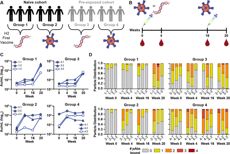
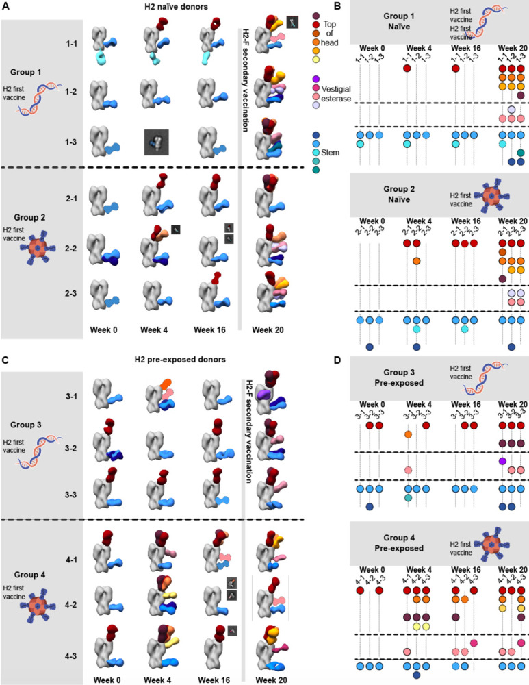
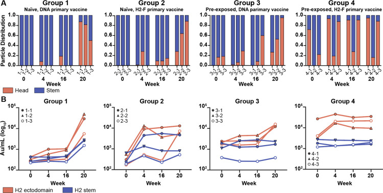
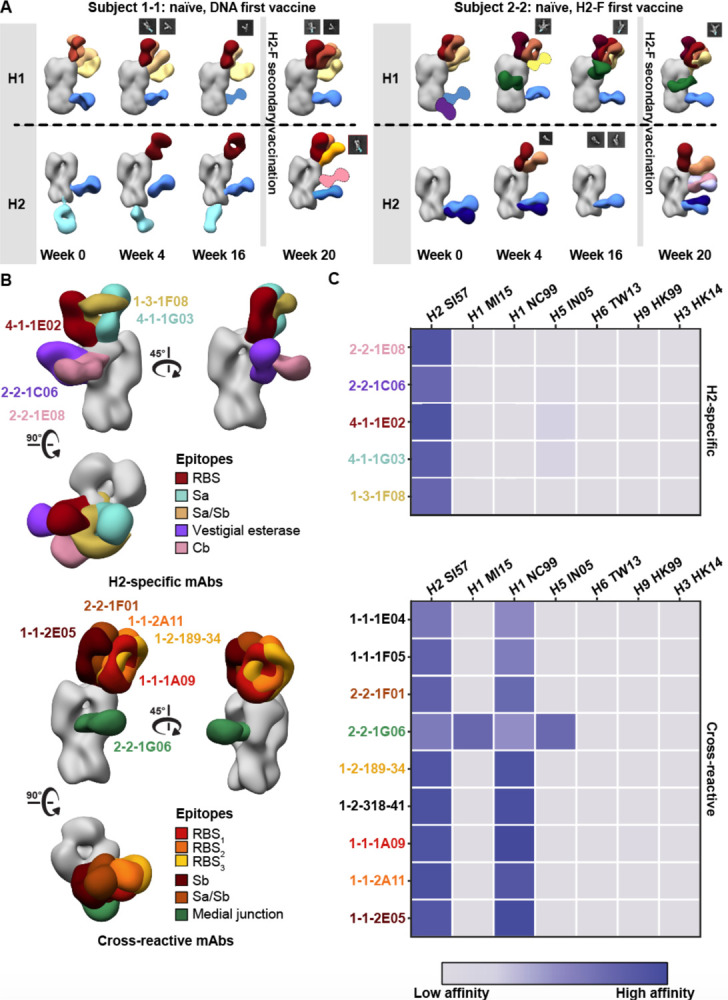
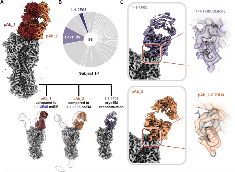
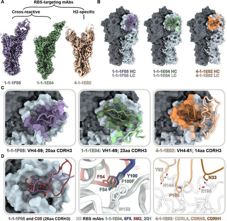
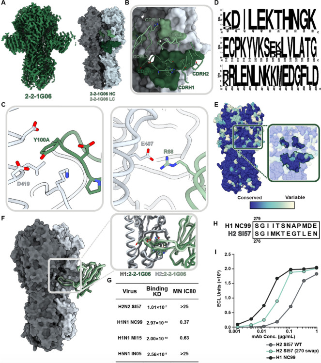
Influenza A virus subtype H2N2, which caused the 1957 influenza pandemic, remains a global threat. A recent phase I clinical trial investigating a ferritin nanoparticle displaying H2 hemagglutinin in H2-naïve and H2-exposed adults. Therefore, we could perform comprehensive structural and biochemical characterization of immune memory on the breadth and diversity of the polyclonal serum antibody response elicited after H2 vaccination. We temporally map the epitopes targeted by serum antibodies after first and second vaccinations and show previous H2 exposure results in higher responses to the variable head domain of hemagglutinin while initial responses in H2-naïve participants are dominated by antibodies targeting conserved epitopes. We use cryo-EM and monoclonal B cell isolation to describe the molecular details of cross-reactive antibodies targeting conserved epitopes on the hemagglutinin head including the receptor binding site and a new site of vulnerability deemed the medial junction. Our findings accentuate the impact of pre-existing influenza exposure on serum antibody responses.

Keywords: cryoEM; hemagglutinin; influenza; neutralizing antibody; structure-based vaccine design.


Conflict of interest statement

DECLARATION OF INTERESTS. The authors declare no conflicting interests.

Figures

Fig. 1.
Fig. 1.. H2N2 vaccine elicits antigen-specific immune responses in trial participants.
(A) Schematic of the H2N2 vaccine trial. Participants were placed into four groups separated by exposure status and vaccination platform: naïve participants who were primed with H2 DNA plasmid-based vaccine (group 1) or the multivalent H2-F nanoparticle (group 2), and pre-exposed participants who were first vaccinated with the DNA plasmid-based vaccine (group 3) or the H2-F nanoparticle (group 4). All groups received secondary vaccinations with H2-F. Individual participants are notated by −1, −2, and −3 for a total of n=3 per group. (B) DNA plasmid and H2-F antigens were administered in two immunizations: first vaccine dose at week 0 and second vaccine dose at week 16. Serum samples were collected at weeks 0, 4, and 16 (after the first vaccination) and week 20 (after boost). (C) MSD binding levels of serum antibodies against H2 HA ectodomain of human participants as measured using Au/ml, arbitrary units/ml. (D) nsEMPEM semi-quantitative epitope occupancy analysis denoting the proportion of HA trimers with 0, 1, 2, 3, or 4 pAbs bound (grey, yellow, orange, dark orange, and red, respectively) for each participant, noted on the x-axis.
Fig. 2.
Fig. 2.. Polyclonal analysis of H2N2 vaccine trial participants.
(A & C) Composite 3D reconstructions with segmented pAb specificities of each participant displayed on one protomer of the H2 HA trimer (grey) for naïve participants (A) or pre-exposed participants (C). Gray lines indicates whether samples were collected pre or post-H2F boost at week 16. Fabs represented as 2D class averages or depicted on the H2 HA trimer as a silhouette with dotted outline have limited particle representation and/or low particle abundance, and their epitopes were consequently predicted. Epitope cluster color scheme is shown on the right. (B & D) Summary of pAb specificities for each group. Each circle represents a unique pAb specificity denoted by the color scheme in A & C.
Fig. 3.
Fig. 3.. Frequency of H2 HA head and stem responses.
(A) nsEMPEM semi-quantitative H2 HA epitope occupancy analysis indicating the proportion of pAb-containing particles in 2D classes targeting the head (orange) or stem (blue). (B) Serum antibody titers measured by MSD using probes of HA ectodomain (orange) and HA stem (blue). Serum samples of each participant are presented with unique symbols.
Fig.4.
Fig.4.. Cross-reactivity of elicited immune responses.
(A) Segmented nsEM 3D reconstructions of participant 1–1 (left) and 2–2 (right) pAbs complexed with either H1/NC99 or H2/1957 HA antigen. Fabs represented as 2D class averages or depicted on the H2 HA trimer as a silhouette with dotted outline have limited particle representation and/or low particle abundance, and their epitopes were consequently predicted. Gray lines indicates whether samples were collected pre- or post-H2F secondary vaccination at week 16. (B) Representative nsEM reconstructions of H2-specific (top) and H1-cross reactive (bottom) monoclonal antibodies in complex with H2 HA. (C) Binding levels of mAbs isolated from plasmablasts or memory B cells against HA subtypes 1 and 2 weeks after H2-F boost.
Fig. 5.
Fig. 5.. Structural analysis of RBS-targeting pAbs in participant 1–1.
(A) CryoEMPEM analysis of immune complexes from participant 1–1 on week 20. H2 HA antigen is colored grey with two segmented Fab density maps colored in red and orange (top). nsEM maps of monoclonal antibodies in complex with H2 HA are overlaid against the corresponding cryo-EM map (bottom). (B) Pie chart showing Ig repertoire of single-cell sorted and sequenced H2-head specific plasmablasts from participant 1–1 one week after the H2HA Ferritin boost. (C) Single-particle cryo-EM reconstruction of H2 HA in complex with 1-1-1F05. (D) Density maps at the epitope-paratope interaction of 1-1-1F05 (top) and pAb_2 (bottom). The atomic model of 1-1-1F05 is shown in purple and docked into both density maps.
Fig. 6.
Fig. 6.. Structural characterization of RBS-targeting antibodies.
(A) CryoEM density maps of mAb-HA complexes. (B) Antibody footprints of 1-1-1F05, 1-1-1E04, and 4-1-1E02 mAbs on HA colored to indicate heavy and light chain interactions. (C) Antibody loop interactions with the RBS pocket, with key CDRH3 residues shown. CDRH3 residue lengths are annotated using the IMGT numbering scheme. (D) 1-1-1F05 and bnAb C05 (PDB 4FP8) CDRH3 loops superimposed (left); 1-1-1E04 superimposed with bnAbs 2G1 (PDB 4HF5) and 8M2 (PDB 4HFU) and 8F8 (PDB 4HF5, middle); and 4-1-1G03 epitope-paratope interaction with key side chains shown (right). (E) Sequence alignment of CDRH3 loops shown in descending order by length.
Fig. 7.
Fig. 7.. Structural and functional characterization of 2-2-1G06 targeting the novel ‘medial junction’ epitope.
(A) CryoEM map of 2-2-1G06 in complex with H2 HA (left) and antibody footprint (right). (B) CDR loop interactions at the 2-2-1G06 epitope. (C) 2-2-1G06 epitope-paratope interactions. Residues presumed critical for binding are shown (Y106 of the CDRH3 on the left and R68 of the CDRL2 on the right). (D) Sequence alignment of 180 human and avian H2 viruses. (E) 16 years of H1 HA sequence variability mapped on an HA surface. Years with sequences represented include 1999, 2006, 2007, 2008, 2009, 2011, 2013, and 2015. (F) Structural comparison of 2-2-1G06 in complex with H2 and H1 NC99. Pop-out panel shows CDRH3 residues. (G) 2-2-1G06 binding affinity and microneutralization of H1, H2, and H5 virus. (H) 270 loop sequence alignment of H1 and H2 strains used in neutralization assay. (I) Binding activity of 2-2-1G06 to SI57 H2 WT, H2 with H1-reverted mutations “270 swap,” and H1 NC99.

References

    1. Lafond K. E. et al. Global burden of influenza-associated lower respiratory tract infections and hospitalizations among adults: A systematic review and meta-analysis. PLoS Med 18, (2021). - PMC - PubMed
    1. Cox N. J. & Subbarao K. GLOBAL EPIDEMIOLOGY OF INFLUENZA: Past and Present*. Annu. Rev. Med vol. 51 www.annualreviews.org (2000). - PubMed
    1. Palese P. Influenza: Old and new threats. Nature Medicine vol. 10 S82–S87 Preprint at 10.1038/nm1141 (2004). - DOI - PubMed
    1. Guan Y. et al. The emergence of pandemic influenza viruses. Protein and Cell vol. 1 9–13 Preprint at 10.1007/s13238-010-0008-z (2010). - DOI - PMC - PubMed
    1. Hobson D., Curry R. L. & Ward-Gardner A. The role of serum haemagglutination-inhibiting antibody in protection against challenge infection with influenza A2 and B viruses. J. Hyg., Camb vol. 70 (1972). - PMC - PubMed

Publication types