

Treatment of Intra-Articular Lesions After Posterior Inferior Tibiofibular Ligament Injury: A Case Series of Elite Rugby Players
- PMID: 37781642
- PMCID: PMC10540585
- DOI: 10.1177/23259671231200934
Treatment of Intra-Articular Lesions After Posterior Inferior Tibiofibular Ligament Injury: A Case Series of Elite Rugby Players
Abstract
Background: Surgical intervention is not typically used to treat symptoms after mild tibiofibular ligament injuries without ankle dislocation or subluxation.
Purpose: To describe outcomes in patients arthroscopically treated for unique intra-articular lesions after sustaining syndesmosis injury of the ankle.
Study design: Case series; Level of evidence, 4.
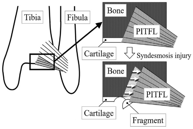
Methods: A total of 11 elite male rugby players with a mean age of 21.0 years (range, 17-28 years) were referred to our hospital for prolonged posterior ankle pain after a high ankle sprain during rugby football. The patients were examined using standing view radiography, computed tomography (CT) and magnetic resonance imaging (MRI) to determine the extent of ligament damage. Posterior ankle arthroscopy was performed to examine intra-articular lesions. The patients were evaluated using the American Orthopaedic Foot and Ankle Society (AOFAS) ankle/hindfoot rating scale and sports activity score of the Self-Administered Foot Evaluation Questionnaire (SAFE-Q).
Results: The average reduced tibiofibular overlap on the standing mortise view was 1.2 mm (range, 0.5-2.0 mm) compared with the opposite ankles. Mason type 1 fracture was detected on CT in 6 patients, and ossification of the interosseous membrane was detected in 2 patients. A bone bruise in the posterior malleolus was observed on MRI in all but 1 patient. Intra-articular fragments located in the posterior ankle were observed and removed arthroscopically. Symptoms improved rapidly after arthroscopic treatment in all patients. All patients returned to rugby games at a median of 11 weeks postoperatively. The median AOFAS scores improved from 77 preoperatively to 100 postoperatively (P < .01), and the median SAFE-Q sports activity subscale score improved from 49.4 to 100 (P < .01).
Conclusion: All unique intra-articular lesions that developed in rugby football players after syndesmosis injury were able to be treated arthroscopically. Patients returned to playing rugby football without syndesmosis reduction. Posterior ankle arthroscopy was effective in patients with residual symptoms after syndesmosis injury.
Keywords: ankle; cartilage; ligament; posterior ankle impingement syndrome; rugby; syndesmosis injury.
© The Author(s) 2023.
Conflict of interest statement
The authors have declared that there are no conflicts of interest in the authorship and publication of this contribution. AOSSM checks author disclosures against the Open Payments Database (OPD). AOSSM has not conducted an independent investigation on the OPD and disclaims any liability or responsibility relating thereto. Ethical approval for this study was obtained from Nara Prefecture General Medical Center (No. 766).
Figures


Similar articles
-
Combination of modified Broström procedure with ankle arthroscopy for chronic ankle instability accompanied by intra-articular symptoms.Arthroscopy. 2010 Apr;26(4):524-8. doi: 10.1016/j.arthro.2010.02.002. Arthroscopy. 2010. PMID: 20362833
-
The prevalence of posterior inferior tibiofibular ligament and inferior tibiofibular transverse ligament injuries in syndesmosis-injured ankles evaluated by oblique axial magnetic resonance imaging: a retrospective study.BMC Musculoskelet Disord. 2022 Mar 18;23(1):264. doi: 10.1186/s12891-022-05220-0. BMC Musculoskelet Disord. 2022. PMID: 35303842 Free PMC article.
-
Anatomical reconstruction of the anterior inferior tibiofibular ligament in elite athletes using InternalBrace suture tape.Bone Joint J. 2022 Jan;104-B(1):68-75. doi: 10.1302/0301-620X.104B1.BJJ-2021-0542.R2. Bone Joint J. 2022. PMID: 34969286
-
Anatomy of the distal tibiofibular syndesmosis in adults: a pictorial essay with a multimodality approach.J Anat. 2010 Dec;217(6):633-45. doi: 10.1111/j.1469-7580.2010.01302.x. J Anat. 2010. PMID: 21108526 Free PMC article. Review.
-
The distal fascicle of the anterior inferior tibiofibular ligament as a cause of tibiotalar impingement syndrome: a current concepts review.Knee Surg Sports Traumatol Arthrosc. 2007 Apr;15(4):465-71. doi: 10.1007/s00167-006-0275-7. Epub 2007 Jan 20. Knee Surg Sports Traumatol Arthrosc. 2007. PMID: 17237964 Free PMC article. Review.
Cited by
-
Safety and clinical efficacy of double posterolateral coaxial portals for endoscopic management of posterior ankle impingement syndrome.Asia Pac J Sports Med Arthrosc Rehabil Technol. 2024 Apr 28;37:8-13. doi: 10.1016/j.asmart.2024.03.006. eCollection 2024 Jul. Asia Pac J Sports Med Arthrosc Rehabil Technol. 2024. PMID: 38706659 Free PMC article.
References
-
- Boggs LR. Isolated posterior malleolar fractures. Am J Emerg Med. 1986;4:334-336. - PubMed
-
- Chang AL, Mandell JC. Syndesmotic ligaments of the ankle: anatomy, multimodality imaging, and patterns of injury. Curr Probl Diagn Radiol. 2020;49(6):452-459. - PubMed
-
- Delahunt E, Farrel G, Boylan A. Mechanism of acute ankle syndesmosis ligament injuries in professional male rugby union players: a systematic visual video analysis. Br J Sports Med. 2021;55(12):691-696. - PubMed
-
- Ferkel RD. Diagnostic arthroscopic examination. In: Ferkel RD, ed. Arthroscopic Surgery: The Foot and Ankle. Lippincott; 1996:103-118.
LinkOut - more resources
Full Text Sources
