. 2023 Nov 28;148(22):1764-1777.
doi: 10.1161/CIRCULATIONAHA.123.065344. Epub 2023 Oct 2.

BRCC3-Mediated NLRP3 Deubiquitylation Promotes Inflammasome Activation and Atherosclerosis in Tet2 Clonal Hematopoiesis

Affiliations

BRCC3-Mediated NLRP3 Deubiquitylation Promotes Inflammasome Activation and Atherosclerosis in Tet2 Clonal Hematopoiesis

Mustafa Yalcinkaya et al. Circulation. .

Abstract

Background: Clonal hematopoiesis (CH) has emerged as an independent risk factor for atherosclerotic cardiovascular disease, with activation of macrophage inflammasomes as a potential underlying mechanism. The NLRP3 (NLR family pyrin domain containing 3) inflammasome has a key role in promoting atherosclerosis in mouse models of Tet2 CH, whereas inhibition of the inflammasome product interleukin-1β appeared to particularly benefit patients with TET2 CH in CANTOS (Cardiovascular Risk Reduction Study [Reduction in Recurrent Major CV Disease Events]). TET2 is an epigenetic modifier that decreases promoter methylation. However, the mechanisms underlying macrophage NLRP3 inflammasome activation in TET2 (Tet methylcytosine dioxygenase 2) deficiency and potential links with epigenetic modifications are poorly understood.

Methods: We used cholesterol-loaded TET2-deficient murine and embryonic stem cell-derived isogenic human macrophages to evaluate mechanisms of NLRP3 inflammasome activation in vitro and hypercholesterolemic Ldlr-/- mice modeling TET2 CH to assess the role of NLRP3 inflammasome activation in atherosclerosis.

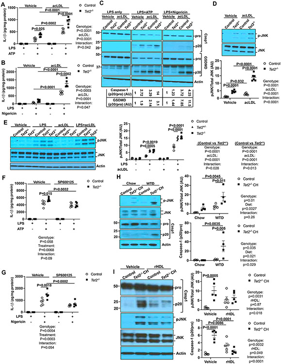
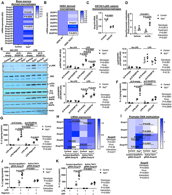
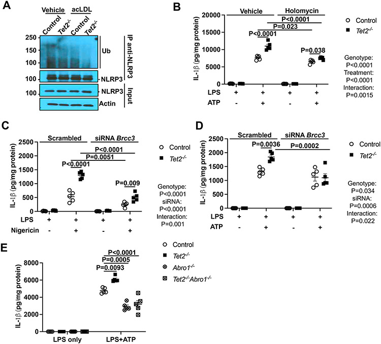
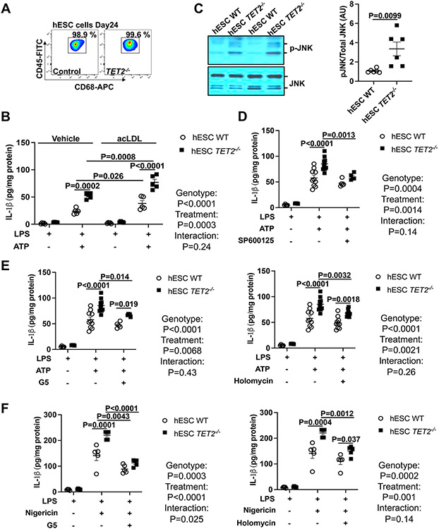
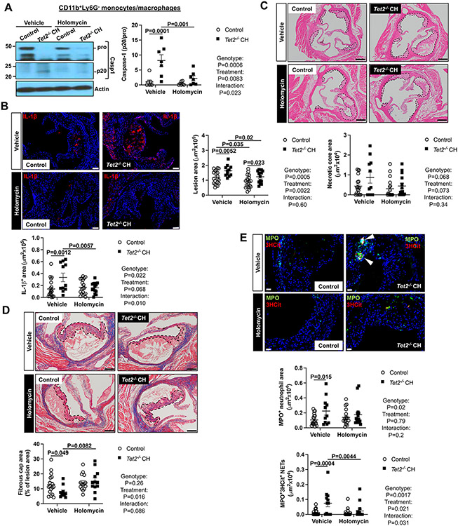
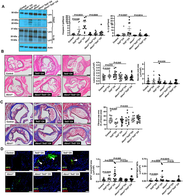
Results: Tet2 deficiency in murine macrophages acted synergistically with cholesterol loading in cell culture and with hypercholesterolemia in vivo to increase JNK1 (c-Jun N-terminal kinase 1) phosphorylation and NLRP3 inflammasome activation. The mechanism of JNK (c-Jun N-terminal kinase) activation in TET2 deficiency was increased promoter methylation and decreased expression of the JNK-inactivating dual-specificity phosphatase Dusp10. Active Tet1-deadCas9-targeted editing of Dusp10 promoter methylation abolished cholesterol-induced inflammasome activation in Tet2-deficient macrophages. Increased JNK1 signaling led to NLRP3 deubiquitylation and activation by the deubiquitinase BRCC3 (BRCA1/BRCA2-containing complex subunit 3). Accelerated atherosclerosis and neutrophil extracellular trap formation (NETosis) in Tet2 CH mice were reversed by holomycin, a BRCC3 deubiquitinase inhibitor, and also by hematopoietic deficiency of Abro1, an essential scaffolding protein in the BRCC3-containing cytosolic complex. Human TET2-/- macrophages displayed increased JNK1 and NLRP3 inflammasome activation, especially after cholesterol loading, with reversal by holomycin treatment, indicating human relevance.

Conclusions: Hypercholesterolemia and TET2 deficiency converge on a common pathway of NLRP3 inflammasome activation mediated by JNK1 activation and BRCC3-mediated NLRP3 deubiquitylation, with potential therapeutic implications for the prevention of cardiovascular disease in TET2 CH.

Keywords: atherosclerosis; clonal hematopoiesis; extracellular traps; hypercholesterolemia; inflammasomes; neutrophils; phosphorylation.


Conflict of interest statement

Disclosures Dr Tall is a consultant for Amgen, CSL Behring, Astra Zeneca, and Foresite Laboratories, and is on the Scientific Advisory Board of Staten Biotech, Fortico Biotech, and Beren Therapeutics.

Figures

Figure 1.
Figure 1.. Elevated JNK activation drives inflammasome activation in Tet2−/− macrophages.
(A-C) Vehicle and acLDL loaded control and Tet2−/− BMDMs were primed with LPS for 3h and treated with ATP or Nigericin for an additional 1h to induce inflammasome activation. (A-B) IL-1β secretion from control and Tet2−/− BMDMs that were treated with ATP (A) or Nigericin (B). (C) Immunoblot of intracellular Caspase-1 and GSDMD cleavage from (A-B). (D) Immunoblot analysis of JNK (Thr183/Tyr185) phosphorylation in vehicle and acLDL loaded control and Tet2−/− BMDMs. (E) Immunoblot of JNK (Thr183/Tyr185) phosphorylation in vehicle and acLDL loaded control, Tet2+/− and Tet2−/− BMDMs upon LPS treatment for 15 min. (F-G) IL-1β from control and Tet2−/− BMDMs that were pretreated with JNK inhibitor SP600125 for 30 min and primed with LPS for 3h and treated with ATP (F) or Nigericin (G) for an additional 1h. (H) Immunoblot analysis of JNK (Thr183/Tyr185) phosphorylation and intracellular Caspase-1 cleavage in Ly6GCD11b+ monocytes/macrophages isolated from Ldlr−/− mice that were transplanted with bone marrow mixture of WT or 10%Tet2−/−/90%WT and fed with chow or WTD for 4 weeks. (I) Immunoblot analysis of JNK (Thr183/Tyr185) phosphorylation and intracellular Caspase-1 cleavage in Ly6GCD11b+ monocytes/macrophages isolated from Ldlr−/− mice that were transplanted with bone marrow mixture of WT or 10%Tet2−/−/90%WT and fed with WTD for 2 weeks and infused with vehicle or reconstituted HDL (rHDL, CSL111) once at the end of the first week. ****P<0.0001, ***P<0.001, ** P<0.01, *P<0.05 by two-way ANOVA with Sidak’s multiple comparison test.
Figure. 2.
Figure. 2.. Dusp10 downregulation due to elevated promoter methylation drives JNK and inflammasome activation in TET2−/− macrophages.
(A) qPCR analysis of dual phosphatases in control and Tet2−/− BMDMs. (B) qPCR analysis of DUSP genes in WT and TET2−/− human embryonic stem cell (hESC)-derived macrophages. (C) Dusp10 mRNA expression in Ly6GCD11b+ monocytes/macrophages isolated from Ldlr−/− mice that were transplanted with bone marrow mixture of WT or 10%Tet2−/−/90%WT and fed with chow or WTD for 4 weeks. (D) meDIP analysis of Dusp10 promoter methylation in vehicle and acLDL loaded control and Tet2−/− BMDMs. (E-G) Control and Tet2−/− BMDMs were infected with control (pLX-empty) or DUSP10 lentiviruses for 72h. (E) Immunoblot analysis of JNK (Thr183/Tyr185), Erk (1/2) and p38 MAPK phosphorylation from cells primed with LPS for 15 min (F) IL-1β secretion from LPS+ATP. (G) IL-1β secretion from LPS+Nigericin. (H-K) Control and Tet2−/− BMDMs were infected with lentiviruses expressing deadCas9-Tet1 (active dC-T) or deadCas9-a catalytically dead form of Tet1 (control dC-dT) with gRNAs targeting the Dusp10 promoter region for 72 hours. (H) qPCR analysis of Dusp genes. (I) meDIP analysis of Dusp promoter methylation. (J) IL-1β secretion from LPS+Nigericin. (K) IL-1β secretion from LPS+ATP. ****P<0.0001, ***P<0.001, ** P<0.01, *P<0.05 by t-test (A-C) and two-way ANOVA with Sidak’s multiple comparison test (D-K).
Figure 3.
Figure 3.. BRCC3-mediated NLRP3 deubiquitylation is essential for inflammasome activation in Tet2 deficiency.
(A) Immunoblot of NLRP3 ubiquitylation in cell lysates immunoprecipitated with anti-NLRP3 in vehicle and acLDL loaded control and Tet2−/− BMDMs primed with LPS for 3h (B) IL-1β secretion from control and Tet2−/− BMDMs were primed with LPS for 3h and treated with holomycin (HL) for 30 min and treated with ATP for additional 1h. (C-D) IL-1β secretion from control and Tet2−/− BMDMs transfected with control (Scrambled) or siRNA against Brcc3 for 48h. After 48 hours of transfection, cells were primed with LPS for 3h and and treated with Nigericin (C) or ATP (D) for an additional 1h. (E) IL-1β secretion from control, Tet2−/−, Abro1−/− and Tet2−/−Abro1−/− BMDMs that were primed with LPS for 3h and treated with ATP for an additional 1h. ****P<0.0001, ***P<0.001, ** P<0.01, *P<0.05 by one-way ANOVA coupled with Tukey’s comparison test (E) and two-way ANOVA with Sidak’s multiple comparison test (B-D).
Figure 4.
Figure 4.. JNK1 mediated NLRP3 deubiquitylation drives NLRP3 inflammasome in human embryonic stem cell-derived TET2−/− macrophages.
(A) The verification of WT and TET2−/− human embryonic stem cell (hESC)-derived macrophages via flow cytometry analysis of CD45 and CD68. (B) IL-1β secretion from WT and TET2−/− hESC-derived macrophages loaded with acLDL overnight and primed with LPS for 3h and treated with ATP for additional 1h. (C) Immunoblot of JNK (Thr183/Tyr185) phosphorylation in WT and TET2−/− hESC-derived macrophages. (D) IL-1β secretion from from WT and TET2−/− hESC-derived macrophages with LPS for 3h in the absence or presence of SP600125 (JNK inhibitor) and treated with ATP for an additional 1h. (E-F) IL-1β secretion from WT and TET2−/− hESC-derived macrophages that were primed with LPS for 3h and treated with Holomycin or G5 for 30 min and treated with ATP (E) or Nigericin (F) for an additional 1h. ****P<0.0001, ***P<0.001, ** P<0.01, *P<0.05 by t-test (C) and two-way ANOVA with Sidak’s multiple comparison test (B, D-F).
Figure 5.
Figure 5.. Inhibition of NLRP3 deubiquitylation decreases NETosis and atherosclerosis in Tet2 CH mice.
Ldlr−/− mice were transplanted with bone marrow mixture of WT or 10%Tet2−/−/90%WT and fed WTD. After 2 weeks WTD feeding, mice were injected intraperitoneally with holomycin (1 mg/kg) or vehicle control (DMSO/PBS) for 6 weeks. Mice were sacrificed after a total of 8 weeks of WTD. (A) Immunoblot of intracellular Caspase-1 cleavage in Ly6GCD11b+splenic monocytes/macrophages. (B) Lesions were stained for IL-1β. (C) Atherosclerotic lesion area and necrotic core area in the aortic root. Representative images of hematoxylin and eosin (H&E)–stained sections are shown; atherosclerotic plaques are delineated by dashed lines. Scale bars, 200 μm. (D) Fibrous cap thickness analysis via Masson trichrome staining. (E) Neutrophils were stained in atherosclerotic lesions using MPO and MPO+ percentages of lesion size was quantified. Concomitantly, lesions were stained for 3HCit. To assess NETs, the overlap of MPO and 3HCit was quantified as percentage of the total lesion area. Representative pictures are shown. Scale bars: 200 μm for (B), 75 μm for (C), 100 μm for (D) and 25 μm for (E). Each datapoint represents an individual mouse. ****P<0.0001, ***P<0.001, ** P<0.01, *P<0.05 by two-way ANOVA with Sidak’s multiple comparison test.
Figure 6.
Figure 6.. Abro1 deficiency decreases NETosis and atherosclerosis in Tet2 CH mice.
Ldlr−/− mice were transplanted with bone marrow mixture WT, 10%Abro1−/−/90%WT, 10%Tet2+/−/90%WT, 10%Tet2−/−/90%WT, 10%Tet2+/−Abro1−/−/90% WT or 10%Tet2−/−Abro1−/−/90% WT. After 5 weeks of recovery time, they were fed with WTD and then were sacrificed after a total of 8 weeks of WTD. (A) Immunoblot of intracellular Caspase-1 and IL-1β cleavage in Ly6GCD11b+splenic monocytes/macrophages. (B) Atherosclerotic lesion area and necrotic core area in the aortic root. Representative images of hematoxylin and eosin (H&E)–stained sections are shown; atherosclerotic plaques are delineated by dashed lines. Scale bars, 200 μm. (C) Fibrous cap thickness analysis via Masson trichrome staining. (D) Neutrophils were stained in atherosclerotic lesions using MPO and MPO+ percentages of lesion size was quantified. Concomitantly, lesions were stained for 3HCit. To assess NETs, the overlap of MPO and 3HCit was quantified as percentage of the total lesion area. Representative pictures are shown. Scale bars: 200 μm for (B), 100 μm for (C) and 50 μm for (D). Each datapoint represents an individual mouse. ****P<0.0001, ***P<0.001, ** P<0.01, *P<0.05 by one-way ANOVA coupled with Tukey’s comparison test for 4 groups (Control, Abro1−/−, Tet2+/−, Tet2+/−Abro1−/−) or (Control, Abro1−/−, Tet2−/− and Tet2−/−Abro1−/−).

References

    1. Ahmad FB, Anderson RN. The Leading Causes of Death in the US for 2020. JAMA. 2021;325:1829–1830. doi: 10.1001/jama.2021.5469 - DOI - PMC - PubMed
    1. Libby P. The changing landscape of atherosclerosis. Nature. 2021;592:524–533. doi: 10.1038/s41586-021-03392-8 - DOI - PubMed
    1. Ridker PM, Everett BM, Thuren T, MacFadyen JG, Chang WH, Ballantyne C, Fonseca F, Nicolau J, Koenig W, Anker SD, et al. Antiinflammatory Therapy with Canakinumab for Atherosclerotic Disease. N Engl J Med. 2017;377:1119–1131. doi: 10.1056/NEJMoa1707914 - DOI - PubMed
    1. Nidorf SM, Fiolet ATL, Mosterd A, Eikelboom JW, Schut A, Opstal TSJ, The SHK, Xu XF, Ireland MA, Lenderink T, et al. Colchicine in Patients with Chronic Coronary Disease. N Engl J Med. 2020;383:1838–1847. doi: 10.1056/NEJMoa2021372 - DOI - PubMed
    1. Tardif JC, Kouz S, Waters DD, Bertrand OF, Diaz R, Maggioni AP, Pinto FJ, Ibrahim R, Gamra H, Kiwan GS, et al. Efficacy and Safety of Low-Dose Colchicine after Myocardial Infarction. N Engl J Med. 2019;381:2497–2505. doi: 10.1056/NEJMoa1912388 - DOI - PubMed

Publication types