. 2023 Dec 1;211(11):1714-1724.
doi: 10.4049/jimmunol.2300053.

Myeloid Heterogeneity Mediates Acute Exacerbations of Pulmonary Fibrosis

Affiliations

Myeloid Heterogeneity Mediates Acute Exacerbations of Pulmonary Fibrosis

Jennifer L Larson-Casey et al. J Immunol. .

Erratum in

Abstract

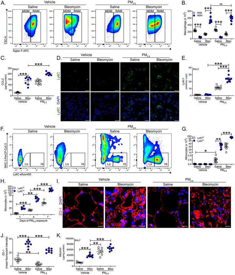
Epidemiological evidence indicates that exposure to particulate matter is linked to the development of idiopathic pulmonary fibrosis (IPF) and increases the incidence of acute exacerbations of IPF. In addition to accelerating the rate of lung function decline, exposure to fine particulate matter (particulate matter smaller than 2.5 μm [PM2.5]) is a risk factor for increased mortality in subjects with IPF. In this article, we show that exposure to PM2.5 mediates monocyte recruitment and fibrotic progression in mice with established fibrosis. In mice with established fibrosis, bronchoalveolar lavage cells showed monocyte/macrophage heterogeneity after exposure to PM2.5. These cells had a significant inflammatory and anti-inflammatory signature. The mixed heterogeneity of cells contributed to the proinflammatory and anti-inflammatory response. Although monocyte-derived macrophages were recruited to the lung in bleomycin-injured mice treated with PM2.5, recruitment of monocytes expressing Ly6Chi to the lung promoted progression of fibrosis, reduced lung aeration on computed tomography, and impacted lung compliance. Ly6Chi monocytes isolated from PM2.5-exposed fibrotic mice showed enhanced expression of proinflammatory markers compared with fibrotic mice exposed to vehicle. Moreover, IPF bronchoalveolar lavage cells treated ex vivo with PM2.5 showed an exaggerated inflammatory response. Targeting Ly6Chi monocyte recruitment inhibited fibrotic progression in mice. Moreover, the adoptive transfer of Ly6Chi monocytes exacerbated established fibrosis. These observations suggest that enhanced recruitment of Ly6Chi monocytes with a proinflammatory phenotype mediates acute exacerbations of pulmonary fibrosis, and targeting these cells may provide a potential novel therapeutic target to protect against acute exacerbations of IPF.


Figures

Figure 1.
Figure 1.. Fibrotic progression occurs in AE-PF
WT mice were exposed to saline or bleomycin (Bleo, 1.75 U/kg i.t.). At day 14, mice were exposed to saline (vehicle) or PM2.5 (8.25 μg in saline, i.t.) daily. Lungs were excised and BAL was performed on day 21. (A) Representative Masson’s trichrome staining of lung tissues. n = 8. Bar = 200 μm. (B) Hydroxyproline analysis of lung homogenates. n = 8. (C) Cell differential using Wright-Giemsa stain to identify mononuclear (mono), neutrophil (PMN), and lymphocytes (lymph). n = 9. (D) Total number of BAL cells. n = 5. (E) Kaplan-Meier survival curve. n = 5. WT mice were exposed to saline or bleomycin. At day 21, mice were exposed to vehicle or PM2.5 daily. Lungs were excised and BAL was performed on day 28. (F) Representative Masson’s trichrome staining of lung tissues. n = 5. Bar = 200 μm. (G) Hydroxyproline analysis of lung homogenates. n = 8-10. (H) Cell differential of BAL cells. n = 9-10. (I) Total number of BAL cells. n = 8-10. (J) Kaplan-Meier survival curve. n = 8. **, p < 0.005; ***, p < 0.0001. Values shown represent means ± S.E.M. One-way ANOVA followed by Tukey’s multiple comparison test was used for B-D, G-I and a log-rank (Mantel-Cox) test was used for E and J. Data shown were pooled from three independent experiments.
Figure 2.
Figure 2.. BAL cells in mice have a mixed phenotype in AE-PF
WT mice were exposed to saline or bleomycin. At day 21, mice were exposed to vehicle or PM2.5 daily. BAL was performed on day 28. mRNA expression for (A) Tnf, (B) Nos2, (C) Tgfb1, and (D) Arg1 in BAL cells. n = 9-10. (E) TNF-α and (F) active TGF-β1 levels in BAL fluid. n = 8-10. IPF BAL cells were obtained by BAL and exposed ex vivo to PM2.5 (10 μg/cm2). mRNA expression of (G) TNF, (H) NOS2, (I) TGFB1, and (J) ARG1 levels in BAL cells. n = 9. *, p < 0.05; ***, p < 0.0001. Values shown represent means ± S.E.M. One-way ANOVA followed by Tukey’s multiple comparison test was used for A-F. Two-tailed t-test statistical analysis was utilized for G-J. Data shown were pooled from three independent experiments.
Figure 3.
Figure 3.. Recruitment of Ly6C expressing monocytes in AE-PF
WT mice were exposed to saline or bleomycin. At day 21, mice were exposed to vehicle or PM2.5 daily. BAL was performed on day 28. (A) Representative flow cytometry plots of resident alveolar macrophages (RAM, CD45+CD11b+/−Ly6GCD64+Ly6CSiglec Fhi) and monocyte-derived macrophages (MDM, CD45+CD11b+/−Ly6GCD64+Ly6CSiglec Flow). (B) Total cell number of RAM and MDM from BAL. n = 9-10. (C) CCL2 levels in BAL fluid. n = 8-10. (D) Representative confocal images of Ly6C positive BAL cells counterstained with DAPI. Bar = 50 μm (n = 5). (E) mRNA expression of Ly6c1 in isolated BAL cells. n = 9-10. (F) Representative flow cytometry plots of Ly6Clo (CD45+CD11b+Ly6GCD64+/−MHC IILy6CloSiglec F) and Ly6Chi (CD45+CD11b+Ly6GCD64+/−MHC IILy6ChiSiglec F) monocytes. (G) Total cell number of Ly6Clo and Ly6Chi monocytes from BAL. n = 9-10. Data shown were pooled from three independent experiments. (H) Number of Ly6Clo and Ly6Chi monocytes from BAL in bleomycin injured mice exposed to PM2.5 for indicated days. n = 6. (I) Representative confocal images of ZO-1 and DAPI staining in mouse lung tissue. Bar = 10 μm (n = 5). (J) Mean fluorescent intensity of ZO-1 staining in lung tissue. n = 8. (K) Albumin levels in BAL fluid. n = 7-8. Data shown are representative from three independent experiments. *, p < 0.05; **, p < 0.001; ***, p < 0.0001. Values shown represent means ± S.E.M. One-way ANOVA followed by Tukey’s multiple comparison test was used. ns = not significant.
Figure 4.
Figure 4.. PM2.5 induces recruitment of inflammatory Ly6Chi monocytes to mediate AE-PF
WT mice were exposed to saline or bleomycin. At day 21, mice were exposed to vehicle or PM2.5 daily. BAL was performed on day 28. (A) Heat map of mRNA expression of FACS-sorted RAMs and MDMs for (B) Tnf, (C) Nos2, (D) Tgfb1, and (E) Arg1. n = 8-10. (F) Heat map of mRNA expression of FACS-sorted Ly6Clo and Ly6Chi monocytes for (G) Tnf, (H) Nos2, (I) Tgfb1, and (J) Arg1. n = 9-10. Data shown were pooled from three independent experiments. (K) Representative micro-CT images of mouse lung parenchyma identifying ≤−500 Hounsfield units (HU, green, aerated lung) and >−500 HU (red, non-aerated lung) using 3D-Slicer imaging software. Quantification of lung density in (L) HU and (M) percentage of aerated lung tissue. n = 6-7. Data shown were pooled from two independent experiments. mtROS generation measured by mitoSOX in (N) isolated BAL cells from exposed mice (n = 6) and (O) FACS-sorted Ly6Chi monocytes from bone marrow exposed to exposed to PM2.5 (10 μg/cm2) and treated with EDTA (5mM). n = 6. Data shown are representative from three independent experiments.*, p < 0.05; **, p < 0.001;***, p < 0.0001. Values shown represent means ± S.E.M. One-way ANOVA followed by Tukey’s multiple comparison test was used. ns = not significant.
Figure 5.
Figure 5.. Ly6Chi monocytes mediate fibrotic progression in mice with advanced fibrosis
(A) Schematic indicating Ly6Chi monocytes derived from bone marrow (1 x 10⁶) were adoptively transferred by i.t. into bleomycin-exposed WT mice 21 days after exposure. (B) Total number of BAL cells. n = 8. mRNA expression in BAL cells for (C) Tnf, (D) Nos2, (E) Tgfb1, and (F) Arg1. n = 7-8. (G) Representative Masson’s trichrome staining of lung tissues. n = 7. Bar = 250 μm. (H) Hydroxyproline analysis of lung homogenates. n = 8. Data shown are representative from two independent experiments. ***, p < 0.0001. Values shown represent means ± S.E.M. Two-tailed t-test statistical analysis was utilized.
Figure 6.
Figure 6.. Ly6Chi monocytes are required for AE-PF
WT mice were exposed to saline or bleomycin. At day 21, mice were exposed to vehicle or PM2.5 daily. On day 23, mice were administered IgG control or anti-CCR2, clone MC-21 antibody (20μg, i.p. daily). BAL was performed on day 28. Representative flow cytometry plots of (A) RAMs and MDMs and (B) Ly6Clo Ly6Chi monocytes. (C) Total cell number of Ly6Clo and Ly6Chi monocytes from BAL. n = 5. mRNA expression for (D) Tnf, (E) Nos2, (F) Tgfb1, and (G) Arg1 in BAL cells. n = 4-5. (H) Representative Masson’s trichrome staining of lung tissues. n = 5. Bar = 250 μm. (I) Hydroxyproline analysis of lung homogenates. n = 8. Data shown were pooled from two independent experiments. (J) Compliance was determined by respiratory mechanics analysis. n = 5-6. **, p < 0.005; ***, p < 0.0001. Values shown represent means ± S.E.M. One-way ANOVA followed by Tukey’s multiple comparison test was used. ns = not significant.

References

    1. Landrigan PJ, Fuller R, Acosta NJR, Adeyi O, Arnold R, Basu NN, Balde AB, Bertollini R, Bose-O'Reilly S, Boufford JI, Breysse PN, Chiles T, Mahidol C, Coll-Seck AM, Cropper ML, Fobil J, Fuster V, Greenstone M, Haines A, Hanrahan D, Hunter D, Khare M, Krupnick A, Lanphear B, Lohani B, Martin K, Mathiasen KV, McTeer MA, Murray CJL, Ndahimananjara JD, Perera F, Potocnik J, Preker AS, Ramesh J, Rockstrom J, Salinas C, Samson LD, Sandilya K, Sly PD, Smith KR, Steiner A, Stewart RB, Suk WA, van Schayck OCP, Yadama GN, Yumkella K, and Zhong M. 2018. The Lancet Commission on pollution and health. Lancet 391: 462–512. - PubMed
    1. Vohra K, Vodonos A, Schwartz J, Marais EA, Sulprizio MP, and Mickley LJ. 2021. Global mortality from outdoor fine particle pollution generated by fossil fuel combustion: Results from GEOS-Chem. Environ. Res 195: 110754. - PubMed
    1. Fann N, Lamson AD, Anenberg SC, Wesson K, Risley D, and Hubbell BJ. 2012. Estimating the national public health burden associated with exposure to ambient PM2.5 and ozone. Risk Anal. 32: 81–95. - PubMed
    1. Laden F, Schwartz J, Speizer FE, and Dockery DW. 2006. Reduction in fine particulate air pollution and mortality: Extended follow-up of the Harvard Six Cities study. Am. J. Respir. Crit. Care Med 173: 667–672. - PMC - PubMed
    1. Harari S, Raghu G, Caminati A, Cruciani M, Franchini M, and Mannucci P. 2020. Fibrotic interstitial lung diseases and air pollution: a systematic literature review. Eur. Respir. Rev 29: 200093. - PMC - PubMed

Publication types

LinkOut - more resources