Ovarian Cancer Patient-Derived Organoid Models for Pre-Clinical Drug Testing
- PMID: 37782106
- PMCID: PMC10881228
- DOI: 10.3791/65068
Ovarian Cancer Patient-Derived Organoid Models for Pre-Clinical Drug Testing
Abstract
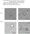
Ovarian cancer is a fatal gynecologic cancer and the fifth leading cause of cancer death among women in the United States. Developing new drug treatments is crucial to advancing healthcare and improving patient outcomes. Organoids are in-vitro three-dimensional multicellular miniature organs. Patient-derived organoid (PDO) models of ovarian cancer may be optimal for drug screening because they more accurately recapitulate tissues of interest than two-dimensional cell culture models and are inexpensive compared to patient-derived xenografts. In addition, ovarian cancer PDOs mimic the variable tumor microenvironment and genetic background typically observed in ovarian cancer. Here, a method is described that can be used to test conventional and novel drugs on PDOs derived from ovarian cancer tissue and ascites. A luminescence-based adenosine triphosphate (ATP) assay is used to measure viability, growth rate, and drug sensitivity. Drug screens in PDOs can be completed in 7-10 days, depending on the rate of organoid formation and drug treatments.
Figures


References
-
- Siegel RL, Miller KD, Jemal A. Cancer statistics, 2019. CA: A Cancer Journal for Clinicians. 69 (1), 7–34 (2019). - PubMed
-
- Christie EL, Bowtell DDL Acquired chemotherapy resistance in ovarian cancer. Annals of Oncology. 28 (suppl_8), viii13–viii15 (2017). - PubMed
-
- Wang L, Wang Q, Xu Y, Cui M, Han L. Advances in the treatment of ovarian cancer using PARP inhibitors and the underlying mechanism of resistance. Current Drug Targets. 21 (2), 167–178 (2020). - PubMed
Publication types
MeSH terms
Grants and funding
LinkOut - more resources
Full Text Sources
Medical
