. 2023 Oct 24;23(21):4664-4673.
doi: 10.1039/d3lc00637a.

A monolithic microfluidic probe for ambient mass spectrometry imaging of biological tissues

Affiliations

A monolithic microfluidic probe for ambient mass spectrometry imaging of biological tissues

Li-Xue Jiang et al. Lab Chip. .

Abstract

Ambient mass spectrometry imaging (MSI) is a powerful technique that allows for the simultaneous mapping of hundreds of molecules in biological samples under atmospheric conditions, requiring minimal sample preparation. We have developed nanospray desorption electrospray ionization (nano-DESI), a liquid extraction-based ambient ionization technique, which has proven to be sensitive and capable of achieving high spatial resolution. We have previously described an integrated microfluidic probe, which simplifies the nano-DESI setup, but is quite difficult to fabricate. Herein, we introduce a facile and scalable strategy for fabricating microfluidic devices for nano-DESI MSI applications. Our approach involves the use of selective laser-assisted etching (SLE) of fused silica to create a monolithic microfluidic probe (SLE-MFP). Unlike the traditional photolithography-based fabrication, SLE eliminates the need for the wafer bonding process and allows for automated, scalable fabrication of the probe. The chamfered design of the sampling port and ESI emitter significantly reduces the amount of polishing required to fine-tune the probe thereby streamlining and simplifying the fabrication process. We have also examined the performance of a V-shaped probe, in which only the sampling port is fabricated using SLE technology. The V-shaped design of the probe is easy to fabricate and provides an opportunity to independently optimize the size and shape of the electrospray emitter. We have evaluated the performance of SLE-MFP by imaging mouse tissue sections. Our results demonstrate that SLE technology enables the fabrication of robust monolithic microfluidic probes for MSI experiments. This development expands the capabilities of nano-DESI MSI and makes the technique more accessible to the broader scientific community.


Conflict of interest statement

There are no conflicts of interest to declare.

Figures

Fig. 1
Fig. 1. Design of the SLE-MFP for nano-DESI MSI: (a) CAD drawing sketching the microfluidic probe design and MS image setup; (b) a photograph of the SLE-MFP in front of the extension tube of a mass spectrometer source inlet; (c) a macro photograph of the monolithic SLE-MFP with the dimensions.
Fig. 2
Fig. 2. (a) Design of the sampling port of an SLE-MFP; photographs of the as-prepared SLE-MFP (b) before and (c) after polishing.
Fig. 3
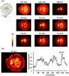
Fig. 3. (a) Optical and representative positive mode ion images of [M + Na]+ ions of phospholipids in a mouse uterine tissue obtained using SLE-MFP. Scale bar: 1 mm; the intensity scale: black (low), yellow (high). The scan rate is 100 μm s−1, and the step between the lines is 60 μm. The acquisition rate of timsTOF is 10 Hz. Ion images are normalized to the internal standard. (b) The spatial resolution of SLE-MFP nano-DESI MSI is determined using the “20–80 rule”. Left: The ion image of [PC 36:4 + Na]+ with the line scan shown in white. Right: Signal profile along the line scan. The steepest gradient used to estimate the spatial resolution is shown with dashed red lines.
Fig. 4
Fig. 4. Design and evaluation of the 2nd generation SLE-MFP with a sharp sampling port for nano-DESI MSI: (a) a schematic diagram of the SLE-MFP with a sharp sampling port; (b) a photograph of the SLE-MFP with a sharp sampling port inlet; (c) optical image and representative positive ion images of molecules in mouse brain tissue obtained using the SLE-MFP. Scale bar: 1 mm; the intensity scale: black (low), yellow (high). The scan rate is 80 μm s−1 and the step between lines is 60 μm. The acquisition rate of MS is 10 Hz. Ion images are normalized to TIC.
Fig. 5
Fig. 5. Design and evaluation of the V-shaped 3rd generation SLE-MFP for nano-DESI MSI: (a) a CAD drawing of the V-shaped SLE-MFP; (b) a photograph of the V-shaped SLE; (c) optical and representative positive mode ion images of [M + Na]+ ions of phospholipids in a mouse uterine tissue obtained using V-shaped SLE-MFP. Scale bar: 1 mm; the intensity scale: black (low), yellow (high). The scan rate is 50 μm s−1 and the step between lines is 50 μm. The ion images are normalized to the internal standard; (d) optical image and representative positive ion images of [M + Na]+ ions of molecules in a mouse brain tissue obtained using the V-shape SLE-MFP. The imaged sample area is marked with a red box as shown in the optical image. The scan rate is 200 μm s−1 and the step between lines is 50 μm. Ion images are normalized to the TIC.

References

    1. Buchberger A. R. DeLaney K. Johnson J. Li L. Anal. Chem. 2018;90:240–265. doi: 10.1021/acs.analchem.7b04733. - DOI - PMC - PubMed
    1. Norris J. L. Caprioli R. M. Chem. Rev. 2013;113:2309–2342. doi: 10.1021/cr3004295. - DOI - PMC - PubMed
    1. McDonnell L. A. Heeren R. M. A. Mass Spectrom. Rev. 2007;26:606–643. doi: 10.1002/mas.20124. - DOI - PubMed
    1. Swales J. G. Hamm G. Clench M. R. Goodwin R. J. A. Int. J. Mass Spectrom. 2019;437:99–112. doi: 10.1016/j.ijms.2018.02.007. - DOI
    1. Schulz S. Becker M. Groseclose M. R. Schadt S. Hopf C. Curr. Opin. Biotechnol. 2019;55:51–59. doi: 10.1016/j.copbio.2018.08.003. - DOI - PubMed

Publication types