. 2023 Oct 10;120(41):e2221985120.
doi: 10.1073/pnas.2221985120. Epub 2023 Oct 2.

Characteristics and anatomic location of PD-1+TCF1+ stem-like CD8 T cells in chronic viral infection and cancer

Affiliations

Characteristics and anatomic location of PD-1+TCF1+ stem-like CD8 T cells in chronic viral infection and cancer

Se Jin Im et al. Proc Natl Acad Sci U S A. .

Abstract

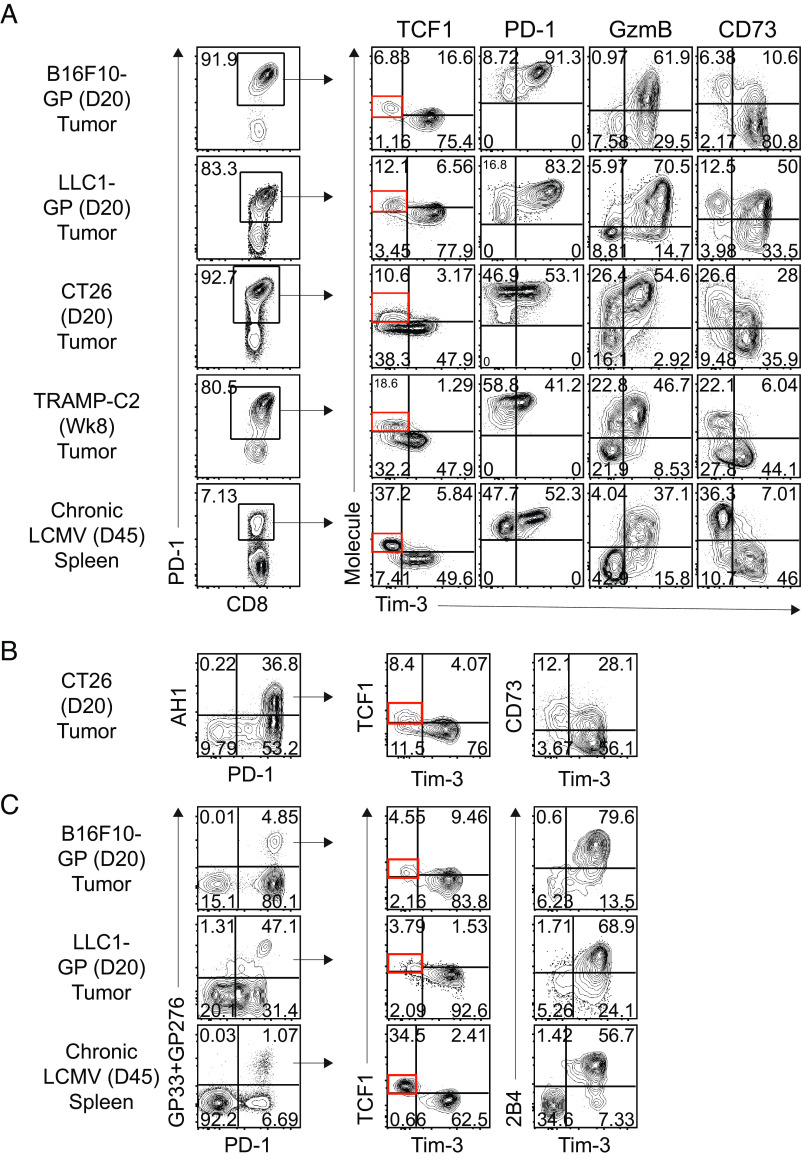
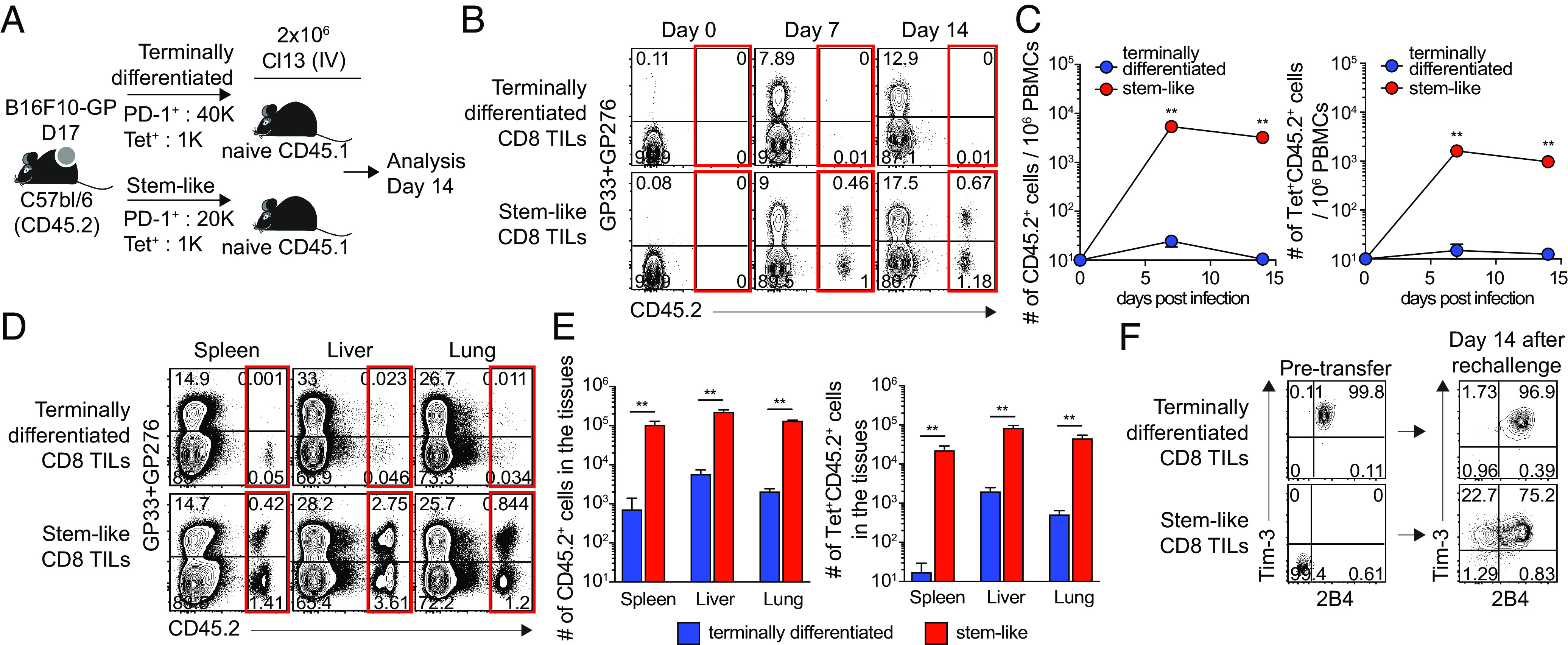
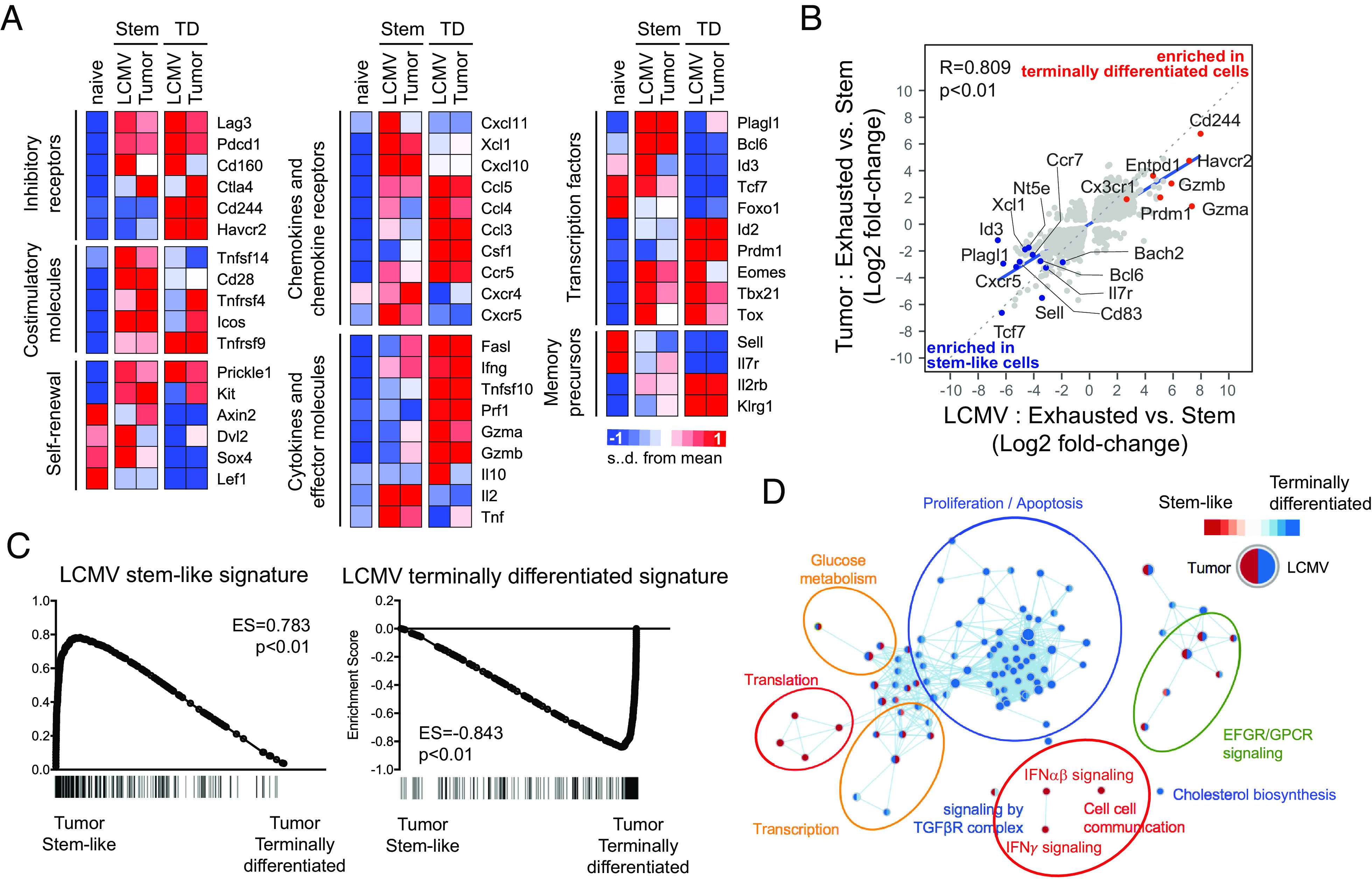
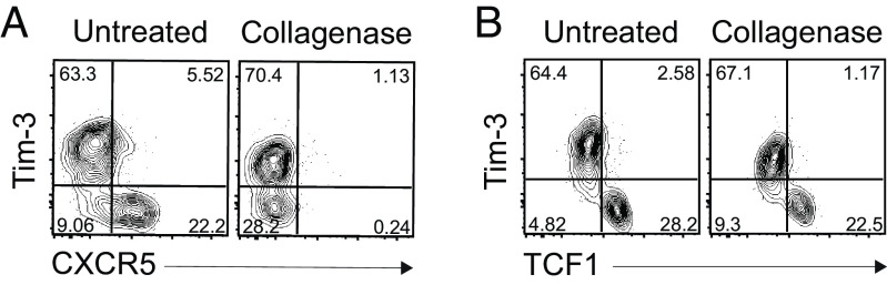
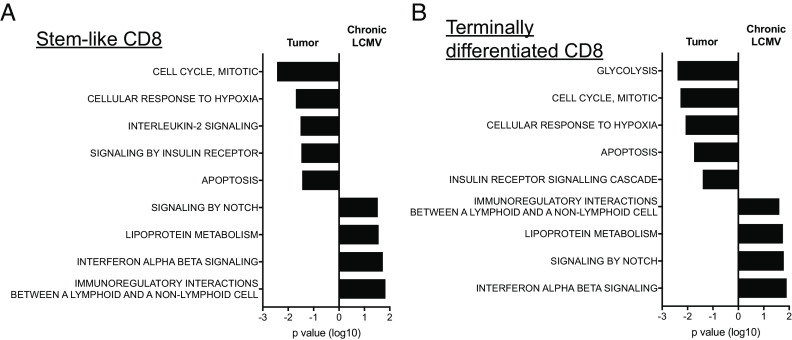
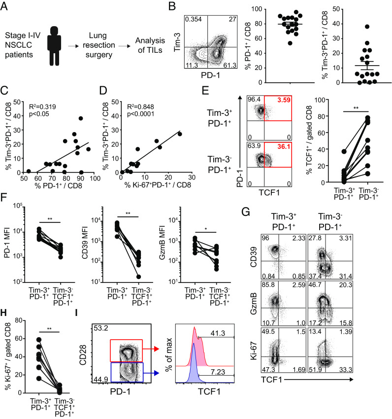
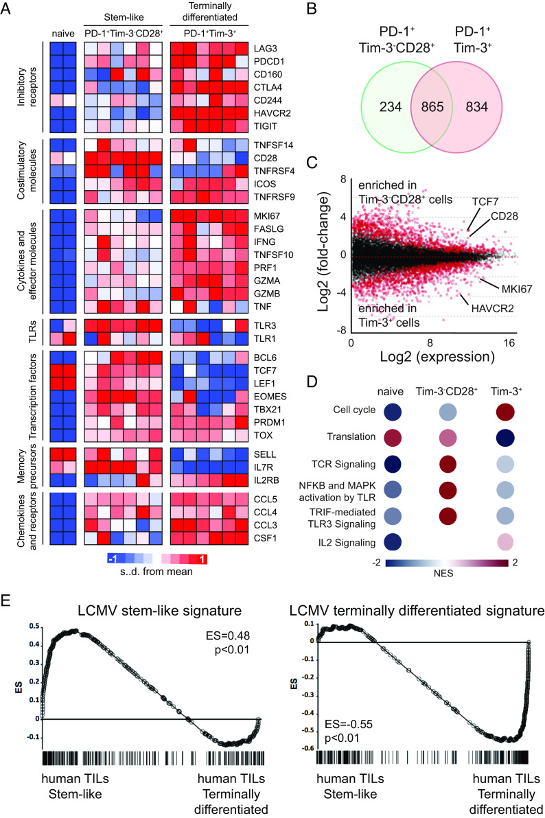
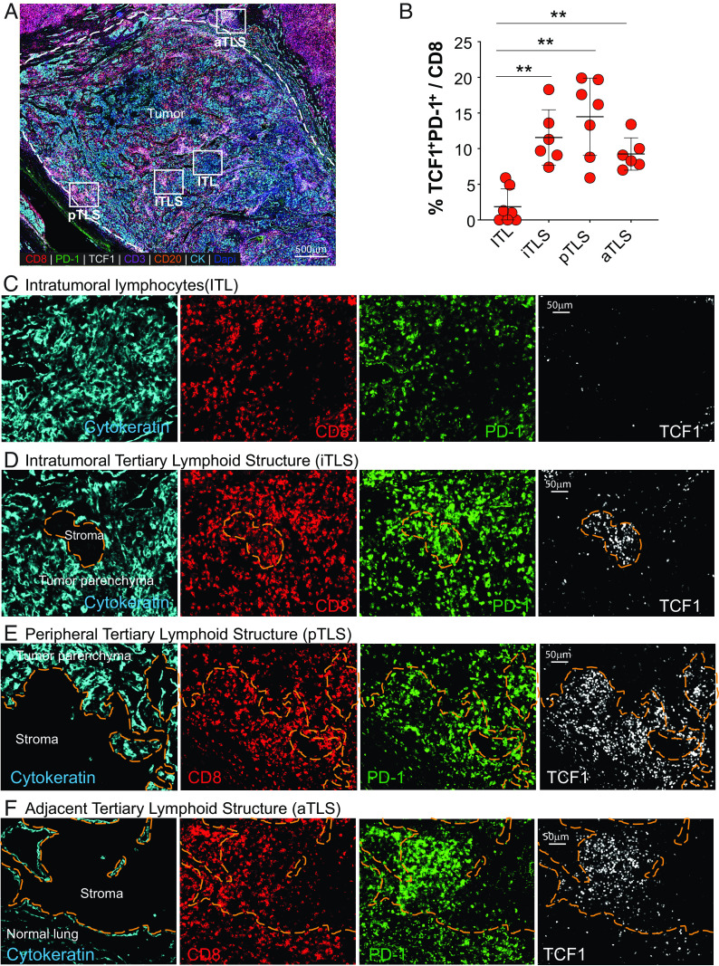
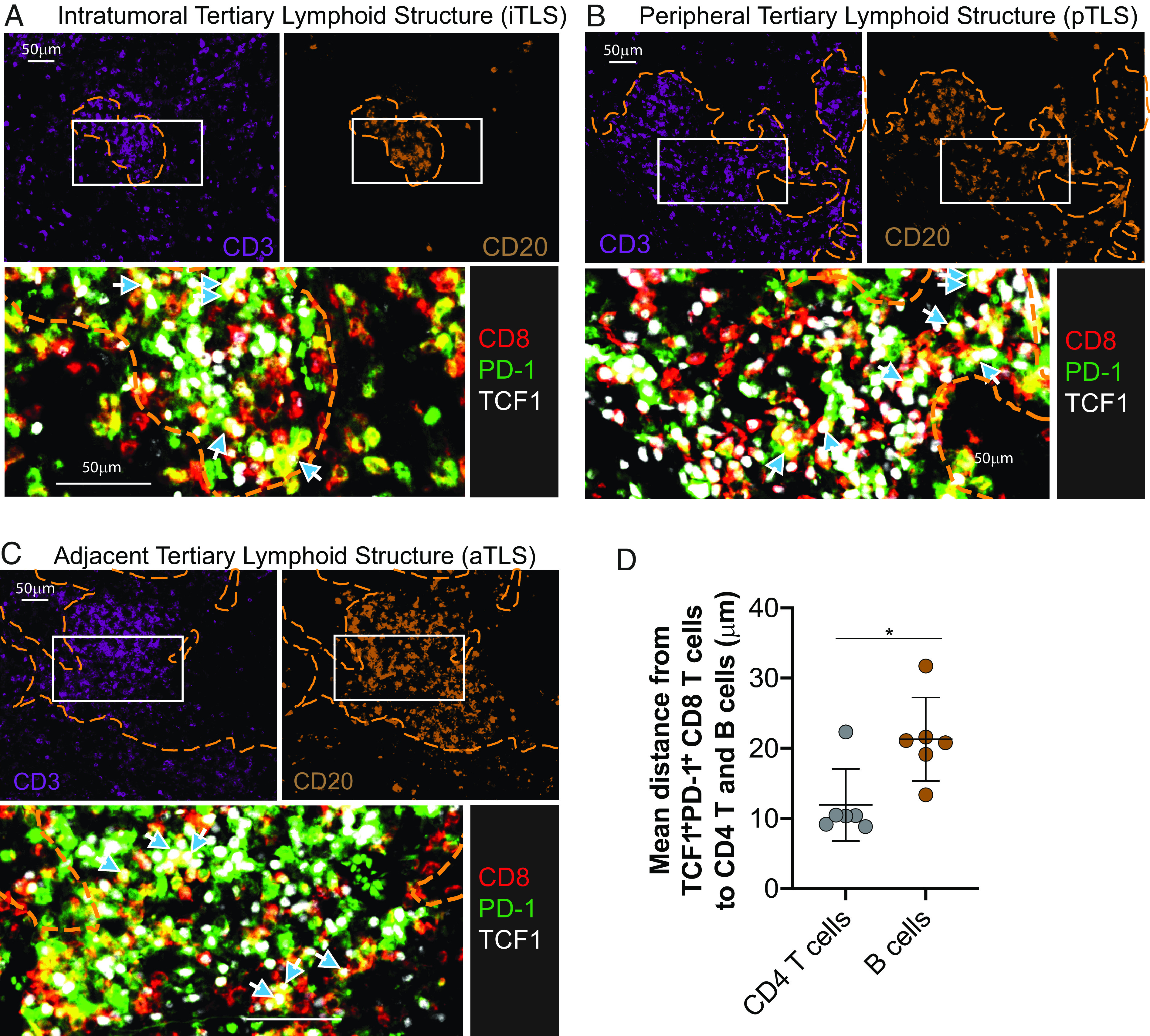
CD8 T cells play an essential role in antitumor immunity and chronic viral infections. Recent findings have delineated the differentiation pathway of CD8 T cells in accordance with the progenitor-progeny relationship of TCF1+ stem-like and Tim-3+TCF1- more differentiated T cells. Here, we investigated the characteristics of stem-like and differentiated CD8 T cells isolated from several murine tumor models and human lung cancer samples in terms of phenotypic and transcriptional features as well as their location compared to virus-specific CD8 T cells in the chronically lymphocytic choriomeningitis virus (LCMV)-infected mice. We found that CD8 tumor-infiltrating lymphocytes (TILs) in both murine and human tumors exhibited overall similar phenotypic and transcriptional characteristics compared to corresponding subsets in the spleen of chronically infected mice. Moreover, stem-like CD8 TILs exclusively responded and produced effector-like progeny CD8 T cells in vivo after antigenic restimulation, confirming their lineage relationship and the proliferative potential of stem-like CD8 TILs. Most importantly, similar to the preferential localization of PD-1+ stem-like CD8 T cells in T cell zones of the spleen during chronic LCMV infection, we found that the PD-1+ stem-like CD8 TILs in lung cancer samples are preferentially located not in the tumor parenchyma but in tertiary lymphoid structures (TLSs). The stem-like CD8 T cells are present in TLSs located within and at the periphery of the tumor, as well as in TLSs closely adjacent to the tumor parenchyma. These findings suggest that TLSs provide a protective niche to support the quiescence and maintenance of stem-like CD8 T cells in the tumor.

Keywords: cancer models; chronic viral infection; human lung cancer; stem-like CD8 T cells; tertiary lymphoid structures.


Conflict of interest statement

The authors declare no competing interest.

Figures

Fig. 1.
Fig. 1.
Identification of stem-like and terminally differentiated CD8 TIL subsets in solid murine tumors. Phenotypic analysis of PD-1+ cells (A) and tumor-specific tetramer-positive CD8 T cells (B and C) in the indicated tumors and the spleen of tumor-bearing mice and chronically LCMV-infected mice, respectively, at the indicated time points. Data are representative of three independent experiments (n = 5/experiment).
Fig. 2.
Fig. 2.
Proliferative potential of stem-like CD8 T cells isolated from tumor-bearing mice upon antigen reexposure. (A) Experimental setup illustrating transfer of congenically marked Tim-3+2B4+ PD-1+ (terminally differentiated) and Tim-32B4 PD-1+ (stem-like) CD8 T cells isolated from B16F10-GP-bearing mice (day 17) into naive Ly5.1 recipient mice, followed by LCMV Cl-13 challenge. (B and C) Representative FACS plots (B) and kinetics of total donor cells and donor GP33/GP276-specific CD8 T cells (C) in the blood (D and E) Representative FACS plots (D) and the number of total donor cells and donor GP33/GP276-specific CD8 T cells (E) in the tissues at day 14 post the challenge. (F) Phenotypic analysis of donor CD8 TIL subsets before and after the transfer followed by Cl-13 challenge. Data were representative of two independent experiments (n = 4/experiment). Graph shows the mean and SEM. Student’s t test, where **P < 0.01.
Fig. 3.
Fig. 3.
Similarities in transcriptional profiles of CD8 T cell subsets between tumor and chronic LCMV infection. Tim-32B4 PD-1+ stem-like and Tim-3+2B4+ PD-1+ terminally differentiated CD8 TILs were isolated from B16F10-GP tumor-bearing mice at day 19 post inoculation. Affymetrix microarray was performed to examine the transcriptome of isolated CD8 TILs, then they were compared with that of two CD8 T cell subsets obtained from mice chronically infected with LCMV (GSE84105) (A) Heat map displaying the relative expression of genes in naive (CD44loCD127+CD62L+) CD8 T cells from uninfected mice, two CD8 TIL subsets from tumor-bearing mice and two CD8 T cell subsets from chronically infected mice. (B) A scatter plot showing differentially expressed genes with twofold cutoff (in addition to the adjusted P-value < 0.05 cutoff) between two CD8 T cell subsets in chronically infected mice (X axis) and tumor-bearing mice (Y axis). (C) GSEA for identifying specific gene signatures of two murine CD8 TIL subsets compared to gene signatures of LCMV stem-like and exhausted CD8 T cells. (D) Cytoscape network analysis for identifying enriched Reactome pathways in each CD8 T cell subset.
Fig. 4.
Fig. 4.
Digestion of CXCR5 molecules on the stem-like CD8 T cells by collagenase treatment. Representative FACS plots of CXCR5 (A) and TCF1 (B) versus Tim-3 expression on GP33-specific CD8 T cells in the spleen of chronically LCMV-infected mice according to the treatment of the collagenase. Data were representative of two independent experiments (n = 4/experiment).
Fig. 5.
Fig. 5.
Differences in transcriptional profiles of CD8 T cell subsets between tumor and chronic LCMV infection. Affymetrix microarray was performed as described in Fig. 3. Enriched Reactome pathways in Tim-32B4 stem-like (A) and Tim-3+2B4+ terminally differentiated CD8 T cells (B) were compared between tumor and chronic LCMV models.
Fig. 6.
Fig. 6.
Phenotypic characterization of TCF1+Tim-3 CD8 TILs in human NSCLC patients. (A) Overview of study design. (B) Representative FACS plots of PD-1 and Tim-3 expression on CD8 TILs and frequency of PD-1+ and Tim-3+PD-1+ CD8 cells among total CD8 TILs in NSCLC tumors. (C and D) Correlation of the frequency of Tim-3+PD-1+ CD8 cells with that of PD-1+ CD8 cells (C) and Ki-67+PD-1+ CD8 cells (D) among total CD8 TILs. (E) Frequency of TCF1+ cells in Tim-3+PD-1+ and Tim-3PD-1+ CD8 TILs. (F and G) MFI of PD-1, CD39, and granzyme B expression (F) in Tim-3+PD-1+ and Tim-3TCF1+PD-1+ CD8 TILs and representative FACS plots including Ki-67 expression (G). (H) Frequency of Ki-67+ cells in Tim-3+PD-1+ and Tim-3TCF1+PD-1+ CD8 TILs. (I) Representative FACS plots of the frequency of TCF1+ cells in CD28+ and CD28 PD-1+ CD8 TILs. Graph shows the mean and SEM. Paired Student’s t test, where **P < 0.01; *P < 0.05.
Fig. 7.
Fig. 7.
Transcriptional profiles of two CD8 TIL subsets isolated from NSCLC patients. Tim-3CD28+ PD-1+ stem-like and Tim-3+ PD-1+ terminally differentiated CD8 TILs were isolated from NSCLC patients. RNA-seq was performed to examine the transcriptome of isolated CD8 TILs. (A) Heat map displaying the relative expression of genes in naive (CCR7+CD45RA+CD58CD95) CD8 T cells from a healthy subject and a NSCLC patient and two CD8 TIL subsets from six NSCLC patients. (B) DEGs in the stem-like and terminally differentiated CD8 T cells compared to naive CD8 T cells. Venn diagram illustrates the overlap in stem-like and exhausted cells among DEGs. (C) Scatter plot of log2 fold-change between stem-like and terminally differentiated CD8 T cells versus mean average read counts. Red indicates genes that were significantly different (adjusted P-value < 0.01). (D) Reactome pathways which were enriched in each CD8 T cell subset compared to other two subsets. NES, normalized enriched score. (E) GSEA for identifying specific gene signatures of two human CD8 TIL subsets compared to gene signatures of LCMV stem-like and terminally differentiated CD8 T cells.
Fig. 8.
Fig. 8.
Spatial location of stem-like CD8 T cells in primary and metastatic human tumors in the lung. (A) Representative area of a pleomorphic carcinoma of the lung. Areas corresponding to the tumor parenchyma containing intratumoral lymphocytes (ITL), tertiary lymphoid structures in the stroma within the tumor bed (iTLS), at the periphery of the tumor (pTLS), and adjacent to the tumor (aTLS) are designated by the white squares. (B) Summary graph showing the number and frequency of TCF1+PD-1+ CD8 T cells in indicated areas in (A) (n = 7). Horizontal line indicates mean and error bars represent SD. Paired Student’s t test, **P < 0.01; *P < 0.05. (CF) Representative areas of ITL, iTLS, pTLS, and aTLS showing the presence and distribution of CD8+, PD-1+, and TCF1+ cells. Orange dashed region marks the stromal region.
Fig. 9.
Fig. 9.
Proximity of CD4 T cells and B cells to stem-like CD8 T cells in tumors. (AC) Location of stem-like CD8 T cells in relation to B cell and CD4 T cell zones within the indicated TLSs from the pleomorphic carcinoma. (D) Summary plot showing the distance from the nearest stem-like CD8 T cells to each CD4 T cell (CD3+/CD8/CD20) or B cell (CD20+) from six different tumors. Graph shows the mean and SD. Paired Student’s t test, where *P < 0.05.

References

    1. Hashimoto M., et al. , CD8 T cell exhaustion in chronic infection and cancer: Opportunities for interventions. Annu. Rev. Med. 69, 301–318 (2018). - PubMed
    1. McLane L. M., Abdel-Hakeem M. S., Wherry E. J., CD8 T cell exhaustion during chronic viral infection and cancer. Annu. Rev. Immunol. 37, 457–495 (2019), 10.1146/annurev-immunol-041015-055318. - DOI - PubMed
    1. Zajac A. J., et al. , Viral immune evasion due to persistence of activated T cells without effector function. J. Exp. Med. 188, 2205–2213 (1998). - PMC - PubMed
    1. Gallimore A., et al. , Induction and exhaustion of lymphocytic choriomeningitis virus-specific cytotoxic T lymphocytes visualized using soluble tetrameric major histocompatibility complex class I-peptide complexes. J. Exp. Med. 187, 1383–1393 (1998). - PMC - PubMed
    1. Barber D. L., et al. , Restoring function in exhausted CD8 T cells during chronic viral infection. Nature 439, 682–687 (2006). - PubMed

Publication types

Substances