

Evaluation of the diagnostic efficacy of liquid-based cytology obtained via percutaneous ultrasound-guided fine-needle aspiration for pancreatic masses: a large tertiary center's 8-year experience
- PMID: 37783929
- PMCID: PMC11797222
- DOI: 10.1007/s00432-023-05438-y
Evaluation of the diagnostic efficacy of liquid-based cytology obtained via percutaneous ultrasound-guided fine-needle aspiration for pancreatic masses: a large tertiary center's 8-year experience
Abstract
Purpose: There were limited data on the diagnostic efficacy of liquid-based cytology (LBC) for pancreatic tissues acquired by percutaneous ultrasound-guided fine-needle aspiration (US-FNA). This study aimed to evaluate the diagnostic value of LBC acquired via percutaneous US-FNA for pancreatic tumors compared with LBC combined with smear cytology (SC).
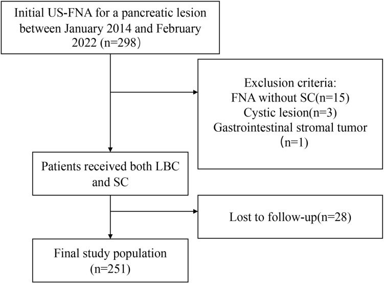
Methods: A retrospective database search (January 2014 and February 2022) was performed for patients who underwent percutaneous US-FNA with both LBC and SC. Clinical and pathological data were collected from 298 patients; eventually, 251 cases met the inclusion criteria. Diagnostic accuracy, sensitivity (SEN), specificity (SPE), positive predictive value (PPV) and negative predictive value (NPV) were compared. Rapid on-site evaluation (ROSE) was not available in all cases.
Results: Based on the pancreaticobiliary cytology guidelines published by the Papanicolaou Society of Cytopathology, 224 (89.2%), 13 (5.2%) and 14 (5.6%) cases were diagnosed as malignant, pre-malignant and benign lesions, respectively. The diagnostic accuracy of the LBC + SC (88.5%) was better than that of LBC (87.3%) but without statistical significance (P = 0.125). The SEN, SPE, PPV and NPV were 87.5%, 85.2%, 98.0% and 45.1%, respectively, in the LBC group and 88.8%, 85.2%, 98.0% and 47.9%, respectively, in the LBC + SC group. According to univariate and multivariate analyses, there were no factors have significant association with the diagnostic sensitivity of LBC.
Conclusions: LBC obtained via percutaneous US-FNA provides good diagnostic value for pancreatic lesions and there was no significant difference between the diagnostic accuracy of LBC and LBC + SC when ROSE was unavailable.
Keywords: Liquid-based cytology; Pancreatic tumor; Ultrasound-guided fine-needle aspiration.
© 2023. The Author(s), under exclusive licence to Springer-Verlag GmbH Germany, part of Springer Nature.
Conflict of interest statement
The authors declare no competing interests.
The authors declare that they have no conflict of interest.
Figures
Similar articles
-
[Health technology assessment report. Use of liquid-based cytology for cervical cancer precursors screening].Epidemiol Prev. 2012 Sep-Oct;36(5 Suppl 2):e1-e33. Epidemiol Prev. 2012. PMID: 23139163 Italian.
-
Imaging modalities for characterising focal pancreatic lesions.Cochrane Database Syst Rev. 2017 Apr 17;4(4):CD010213. doi: 10.1002/14651858.CD010213.pub2. Cochrane Database Syst Rev. 2017. PMID: 28415140 Free PMC article.
-
Treatments for breast abscesses in breastfeeding women.Cochrane Database Syst Rev. 2015 Aug 17;2015(8):CD010490. doi: 10.1002/14651858.CD010490.pub2. Cochrane Database Syst Rev. 2015. PMID: 26279276 Free PMC article.
-
Comparison of smear cytology and liquid-based cytology in EUS-guided FNA of pancreatic lesions: experience from a large tertiary center.Gastrointest Endosc. 2020 Apr;91(4):932-942. doi: 10.1016/j.gie.2019.10.033. Epub 2019 Nov 16. Gastrointest Endosc. 2020. PMID: 31738926
-
EUS FNAC without rapid on-site evaluation is comparable to EUS FNB with macroscopic on-site evaluation in evaluation of intra-abdominal masses.Indian J Gastroenterol. 2025 Jun;44(3):371-377. doi: 10.1007/s12664-025-01741-3. Epub 2025 Feb 19. Indian J Gastroenterol. 2025. PMID: 39969684
References
-
- Bernardes C, Carvalho D, Ramos G (2017) Liquid-based cytology for the diagnosis of solid pancreatic tumors with endoscopic ultrasound-guided fine-needle aspiration: can we dismiss conventional smear? Dig Endosc 29:814. 10.1111/den.12915 - PubMed
-
- Beyer G, Habtezion A, Werner J et al (2020) Chronic pancreatitis. Lancet 396:499–512. 10.1016/s0140-6736(20)31318-0 - PubMed
-
- Chai WL, Kuang XF, Yu L et al (2022) Percutaneous ultrasound and endoscopic ultrasound-guided biopsy of solid pancreatic lesions: an analysis of 1074 lesions. Hepatobiliary Pancreat Dis Int. 10.1016/j.hbpd.2022.06.017 - PubMed
-
- Chun JW, Lee K, Lee SH et al (2020) Comparison of liquid-based cytology with conventional smear cytology for EUS-guided FNA of solid pancreatic masses: a prospective randomized noninferiority study. Gastrointest Endosc 91:837-846.e831. 10.1016/j.gie.2019.11.018 - PubMed
-
- D’Onofrio M, De Robertis R, Barbi E et al (2016) Ultrasound-guided percutaneous fine-needle aspiration of solid pancreatic neoplasms: 10-year experience with more than 2,000 cases and a review of the literature. Eur Radiol 26:1801–1807. 10.1007/s00330-015-4003-x - PubMed
MeSH terms
Grants and funding
LinkOut - more resources
Full Text Sources
Medical
Miscellaneous
