. 2023 Oct 2;54(1):86.
doi: 10.1186/s13567-023-01217-2.

Binding of Trichinella spiralis C-type lectin with syndecan-1 on intestinal epithelial cells mediates larval invasion of intestinal epithelium

Affiliations

Binding of Trichinella spiralis C-type lectin with syndecan-1 on intestinal epithelial cells mediates larval invasion of intestinal epithelium

Zhen Wang et al. Vet Res. .

Abstract

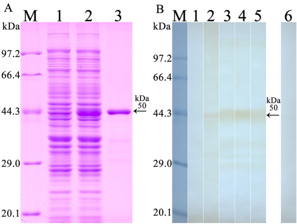
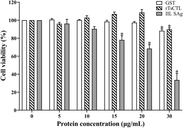
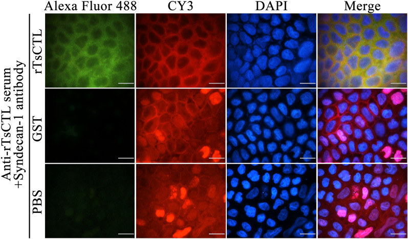
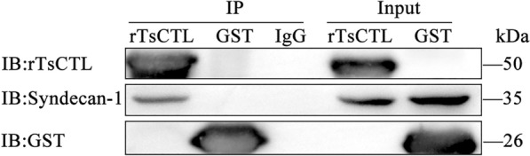
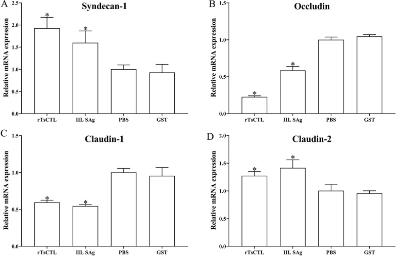
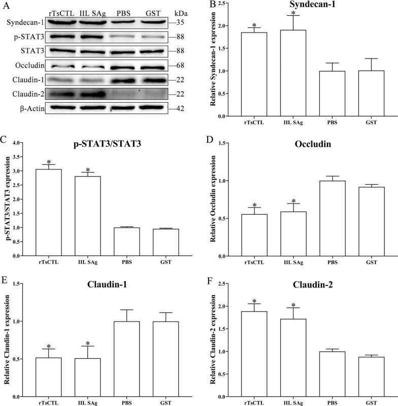
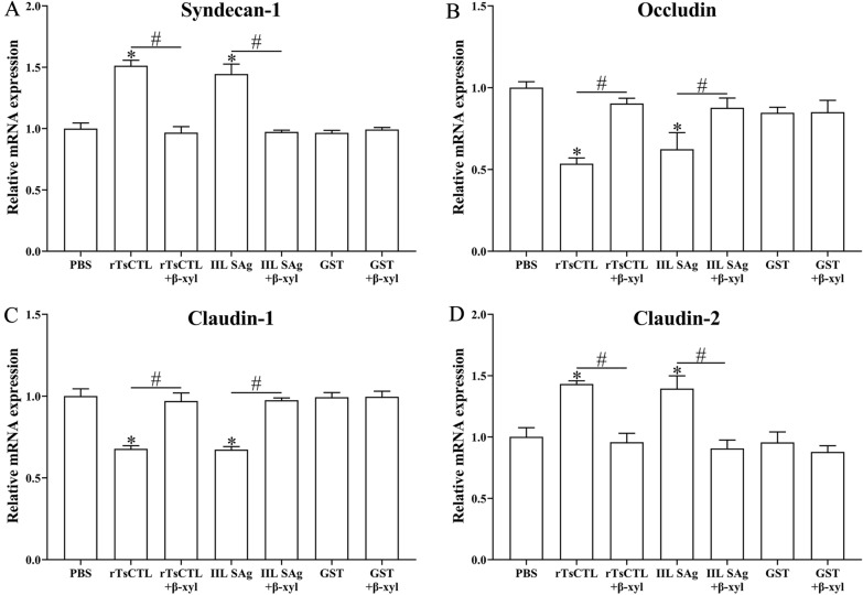
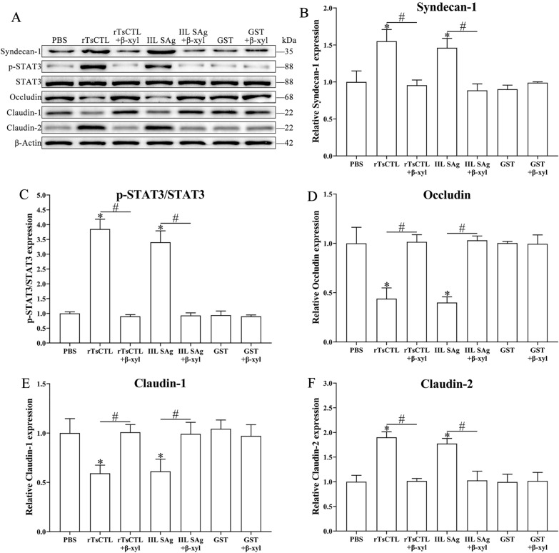
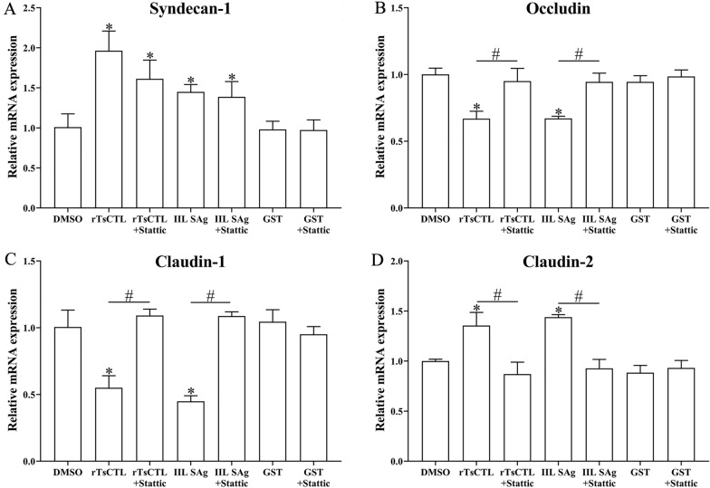
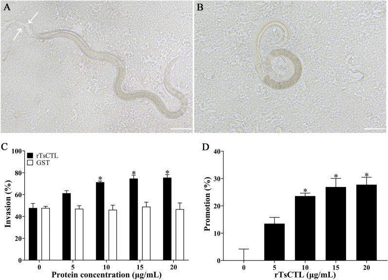
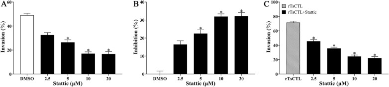
C-type lectin (CTL) is a protein that binds to saccharides and plays an important role in parasite adhesion, host cell invasion and immune evasion. Previous studies showed that recombinant T. spiralis C-type lectin (rTsCTL) promotes larval invasion of intestinal epithelium cells (IEC), whereas anti-rTsCTL antibodies inhibits larval invasion. Syndecan-1 (SDC-1) is a member of the heparan sulfate proteoglycan family which is mainly expressed on the surface of IEC and in extracellular matrices where they interact with a plethora of ligands. SDC-1 has a principal role in maintaining cell morphogenesis, establishing cell-cell adhesions, and regulating the gut mucosal barrier. The aim of this study was to investigate whether rTsCTL binds to SDC-1 on IEC, and the binding of rTsCTL with SDC-1 promotes larval invasion and its mechanism. IFA results show that rTsCTL and SDC-1 co-localized on Caco-2 cell membrane. GST pull-down and Co-IP verified the direct interaction between rTsCTL and SDC-1 on Caco-2 cells. qPCR and Western blotting revealed that rTsCTL binding to SDC-1 increased the expression of SDC-1 and claudin-2, and reduced the expression of occludin and claudin-1 in Caco-2 cells incubated with rTsCTL via the STAT3 pathway. β-Xyloside (a syndecan-1 synthesis inhibitor) and Stattic (a STAT3 inhibitor) significantly inhibited rTsCTL binding to syndecan-1 in Caco-2 cells and activation of the STAT3 pathway, abrogated the effects of rTsCTL on the expression of gut tight junctions, and impeded larval invasion. The results demonstrate that binding of rTsCTL to SDC-1 on Caco-2 cells activated the STAT3 pathway, decreased gut tight junction expression, damaged the integrity of the gut epithelial barrier, and mediated T. spiralis invasion of the gut mucosa. TsCTL might be regarded as a candidate vaccine target against T. spiralis invasion and infection.

Keywords: C-type lectin; Trichinella spiralis; intestinal epithelium; invasion; syndecan-1.


Conflict of interest statement

The authors declare that they have no competing interests.

Figures

Figure 1
Figure 1
Identification of rTsCTL. A SDS-PAGE analysis of rTsCTL. Lane M: protein marker; Lane 1: lysate of bacteria carrying pGEX-4T-1/TsCTL prior to induction; Lane 2: lysate of bacteria carrying pGEX-4T-1/TsCTL after induction; Lane 3: purified rTsCTL (black arrow). B Western blotting analysis of rTsCTL. The lysates of bacteria carrying pGEX-4T-1/TsCTL prior to induction (lane 1) was not recognized by infection serum; lysate of induced bacteria carrying pGEX-4T-1/TsCTL (lane 2), purified rTsCTL (lane 3–6) were recognized with infection serum (lane 2, 3), anti-rTsCTL serum (lane 4) and anti-GST-tag serum (lane 5), but not by normal serum (lane 6).
Figure 2
Figure 2
The effect of rTsCTL, IIL SAg and GST on the viability of Caco-2 cells. Caco-2 cells were incubated with different concentrations of GST, rTsCTL and IIL SAg for 24 h, and cell viability was detected. Cell viability = (OD values of test group − OD values of blank control)/(OD values of GST tag control − OD values of blank control) × 100%. *P < 0.001 indicates an obvious reduction of cell activity compared to the blank control group.
Figure 3
Figure 3
Immunofluorescence co-localization of rTsCTL and syndecan-1. Alexa Fluor 488: Caco-2 cells were incubated with rTsCTL, GST and PBS, then probed by anti-rTsCTL serum, and stained by Alexa Fluor 488-conjugated anti-mouse IgG. CY3: Caco-2 cells were probed by anti-syndecan-1 antibody, and stained by CY3-conjugated anti-rabbit IgG. DAPI: cell nuclei were stained by DAPI as blue; scale bars: 40 μm.
Figure 4
Figure 4
Binding of rTsCTL to syndecan-1 in Caco-2 cells assessed by GST pull-down and Western blot analysis. Lane 1: GST resins + rTsCTL; Lane 2: GST resins + rTsCTL + Caco-2 cell proteins; Lane 3: GST resins + GST; Lane 4: GST resins + GST + Caco-2 cell proteins; Lane 5: The lysates of Caco-2 cells; Lane 6: GST resins + Caco-2 cell proteins.
Figure 5
Figure 5
Binding of rTsCTL and syndecan-1 in Caco-2 cells detected by Co-IP. rTsCTL: IP samples of Caco-2 cellular protein incubated with anti-rTsCTL serum; GST: IP samples of Caco-2 cell proteins incubated with anti-GST serum; IgG: IP samples of Caco-2 cell proteins incubated with normal mouse IgG; Input: input rTsCTL, Caco-2 cell lysate and GST were examined by using anti-rTsCTL serum, anti-syndecan-1 antibody and anti-GST serum; IP: immunoprecipitation; IB: immunoblotting.
Figure 6
Figure 6
qPCR analysis of the transcription levels of syndecan-1 and TJs in Caco-2 cells incubated with rTsCTL. Caco-2 cells were incubated with rTsCTL (5 µg/mL), and IIL SAg and GST tag protein were respectively used as a positive or negative control. The transcription levels of syndecan-1 (A), occludin (B), claudin-1 (C), and claudin-2 (D) were analyzed by qPCR. The transcription levels were calculated with the 2−ΔΔCt method. β-Actin was used as an internal control. *P < 0.05 compared to the PBS group.
Figure 7
Figure 7
Western blotting of expression levels of syndecan-1, p-STAT3 and TJs in Caco-2 cells incubated with rTsCTL. A Caco-2 cells were incubated with rTsCTL (5 µg/mL), and IIL SAg and GST tag protein were respectively used as a positive or negative control. The expression levels of syndecan-1, p-STAT3, STAT3, occludin, claudin-1, and claudin-2 were analyzed by Western blotting, and β-Actin was used as an internal reference control. BF Densitometric analysis of protein bands obtained in panel (A) for syndecan-1 (B), p-STAT3/STAT3 (C), occludin (D), claudin-1 (E) and claudin-2 (F) relative to the β-Actin band *P < 0.01 compared to the PBS group.
Figure 8
Figure 8
qPCR analysis of transcription levels of syndecan-1 and TJs in Caco-2 cells after β-xyloside treatment. Caco-2 cells were pretreated with β-xyloside (5 mM) and then incubated with rTsCTL (5 µg/mL), and IIL SAg and GST tag protein were respectively used as a positive or negative control. The transcription levels of syndecan-1 (A), occludin (B), claudin-1 (C), and claudin-2 (D) were ascertained by qPCR. The transcription levels were calculated with the 2−ΔΔCt method. β-Actin was used as an internal control. *P < 0.01 compared to the PBS group. #P < 0.001 compared between two groups.
Figure 9
Figure 9
Western blot analysis of expression levels of syndecan-1, p-STAT3 and TJs in Caco-2 cells after β-xyloside treatment. A Caco-2 cells were pretreated with β-xyloside (5 mM) and then incubated with rTsCTL (5 µg/mL), and IIL SAg and GST tag protein were respectively used as a positive or negative control. The expression levels of syndecan-1, p-STAT3, STAT3, occludin, claudin-1, and claudin-2 were analyzed by Western blot, and β-Actin was used as an internal reference control. BF Densitometric analysis of the bands obtained in (A) panel for syndecan-1 (B), p-STAT3/STAT3 (C), occludin (D), claudin-1 (E) and claudin-2 (F) relative to the β-Actin band *P < 0.01 compared with PBS. #P < 0.01 compared between two groups.
Figure 10
Figure 10
qPCR analysis of transcription levels of syndecan-1 and TJs in Caco-2 cells treated with Stattic. Caco-2 cells were pre-treated with Stattic (10 µM) and then incubated with rTsCTL (5 µg/mL), 0.1% DMSO (Stattic solvent) was used as a negative control. The transcription levels of syndecan-1 (A), occludin (B), claudin-1 (C), and claudin-2 (D) were assessed by qPCR. The transcription levels were calculated with the 2−ΔΔCt method. β-Actin was used as an internal control. *P < 0.05 compared to the DMSO group. #P < 0.05 compared between two groups.
Figure 11
Figure 11
Western blotting analysis of expression levels of syndecan-1, p-STAT3 and TJ in Caco-2 cells after Stattic treatment. A Caco-2 cells were pre-treated with Stattic (10 µM) and then incubated with rTsCTL (5 µg/mL), and 0.1% DMSO (Stattic solvent) was used as a negative control. The expression levels of syndecan-1, p-STAT3, STAT3, occludin, claudin-1, and claudin-2 were ascertained by Western blotting, and β-Actin was used as an internal reference control. BF Densitometric analysis of the bands obtained in (A) for syndecan-1 (B), p-STAT3/STAT3 (C), occludin (D), claudin-1 (E) and claudin-2 (F) relative to the β-Actin band. *P < 0.01 relative to the DMSO group. #P < 0.01 compared between two groups.
Figure 12
Figure 12
Facilitation of rTsCTL on larval invasion of Caco-2 cells. A The invaded larva was mobile and migratory in the monolayer (the white arrows showed the migratory trace). B Non-invaded larva was coiled on the Caco-2 surface. C and D rTsCTL accelerated IIL invasion into Caco-2 cells. Scale bars: 100 μm. *P < 0.05 compared to the GST or PBS control group. Promotion (%) = Invasion rate of the experimental group − average invasion rate of the PBS control group.
Figure 13
Figure 13
β-xyloside inhibited larval invasion of Caco-2 cells and abrogated rTsCTL facilitative role on the invasion. A and B 10 and 20 mM β-xyloside significantly inhibited larval invasion of Caco-2 cells. C 2.5–20 mM β-xyloside significantly inhibited and abrogated the rTsCTL promotion role on larval invasion of Caco-2 cells. * P < 0.05 compared to the PBS group or only rTsCTL group. Inhibition (%) = average invasion rate of the PBS control group − invasion rate of the experimental group.
Figure 14
Figure 14
Stattic inhibited larval invasion of Caco-2 cells and abrogated rTsCTL facilitative role on the invasion. A and B Stattic at 5, 10 and 20 µM significantly inhibited larval invasion of Caco-2 cells. C 2.5–20 µM Stattic significantly inhibited and abrogated the rTsCTL promotion role on the in vitro larval invasion of Caco-2 cells. *P < 0.05 compared to the DMSO or only rTsCTL group. Inhibition (%) = Average invasion rate of the DMSO control group − invasion rate of the experimental group.

References

    1. Pozio E. The broad spectrum of Trichinella hosts: from cold- to warm-blooded animals. Vet Parasitol. 2005;132:3–11. - PubMed
    1. Rostami A, Gamble HR, Dupouy-Camet J, Khazan H, Bruschi F. Meat sources of infection for outbreaks of human trichinellosis. Food Microbiol. 2017;64:65–71. - PubMed
    1. Zhang XZ, Wang ZQ, Cui J. Epidemiology of trichinellosis in the people’s Republic of China during 2009–2020. Acta Trop. 2022;229:106388. - PubMed
    1. Bai SJ, Han LL, Liu RD, Long SR, Zhang X, Cui J, Wang ZQ. Oral vaccination of mice with attenuated Salmonella encoding Trichinella spiralis calreticulin and serine protease 1.1 confers protective immunity in BALB/c mice. PLoS Negl Trop Dis. 2022;16:e0010929. - PMC - PubMed
    1. Tang B, Li J, Li T, Xie Y, Guan W, Zhao Y, Yang S, Liu M, Xu D. Vaccines as a strategy to control trichinellosis. Front Microbiol. 2022;13:857786. - PMC - PubMed