. 2023 Oct 2;11(1):159.
doi: 10.1186/s40478-023-01647-1.

Higher angiotensin-converting enzyme 2 (ACE2) levels in the brain of individuals with Alzheimer's disease

Affiliations

Higher angiotensin-converting enzyme 2 (ACE2) levels in the brain of individuals with Alzheimer's disease

Louise Reveret et al. Acta Neuropathol Commun. .

Erratum in

Abstract

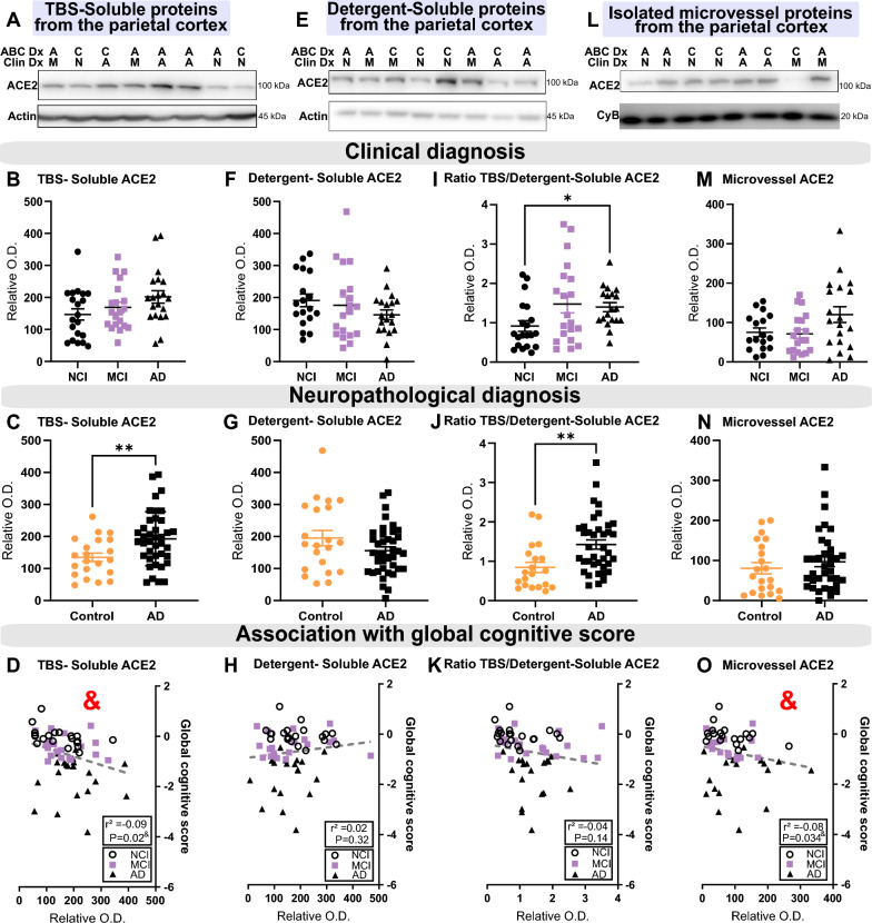
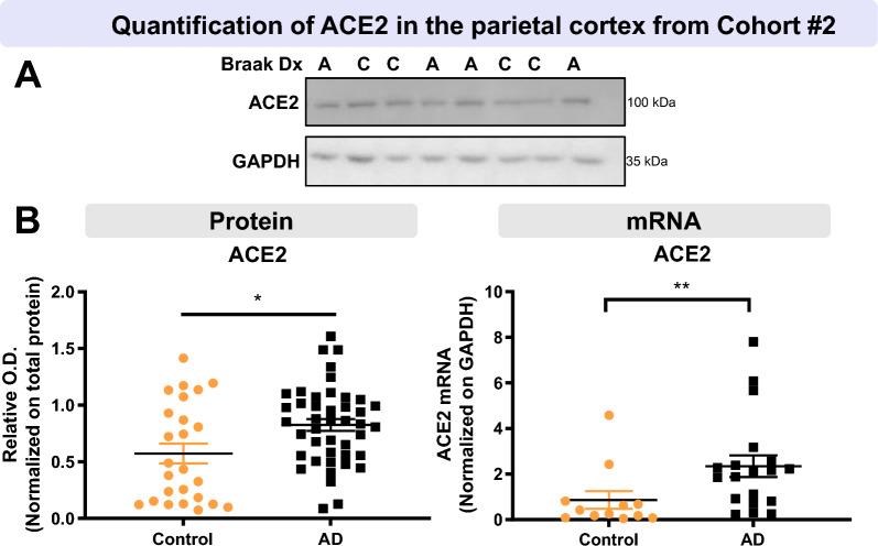
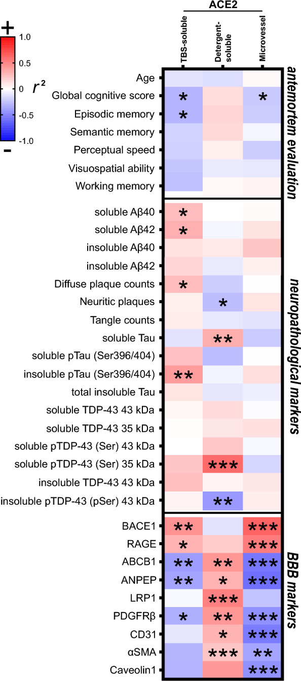
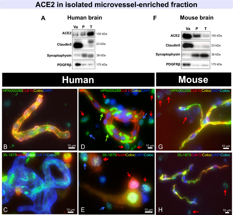
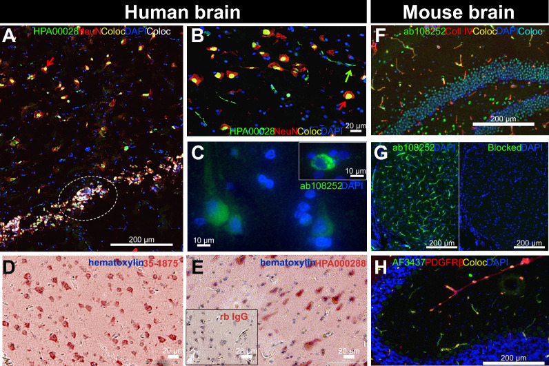
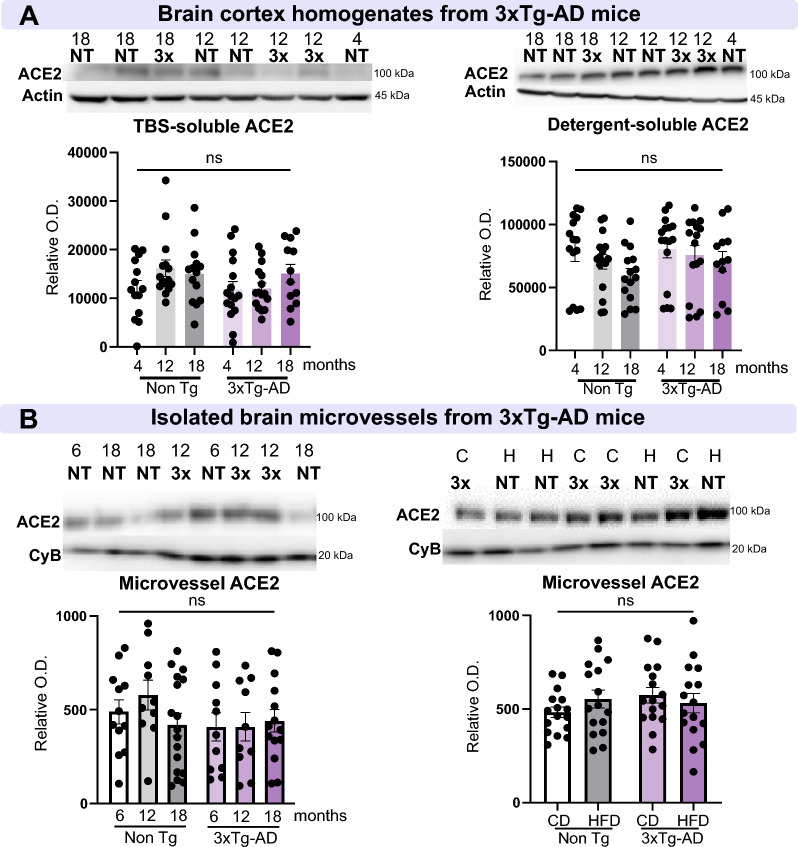
Cognitive decline due to Alzheimer's disease (AD) is frequent in the geriatric population, which has been disproportionately affected by the COVID-19 pandemic. In this study, we investigated the levels of angiotensin-converting enzyme 2 (ACE2), a regulator of the renin-angiotensin system and the main entry receptor of SARS-CoV-2 in host cells, in postmortem parietal cortex samples from two independent AD cohorts, totalling 142 persons. Higher concentrations of ACE2 protein (p < 0.01) and mRNA (p < 0.01) were found in individuals with a neuropathological diagnosis of AD compared to age-matched healthy control subjects. Brain levels of soluble ACE2 were inversely associated with cognitive scores (p = 0.02) and markers of pericytes (PDGFRβ, p = 0.02 and ANPEP, p = 0.007), but positively correlated with concentrations of soluble amyloid-β peptides (Aβ) (p = 0.01) and insoluble phospho-tau (S396/404, p = 0.002). However, no significant differences in ACE2 were observed in the 3xTg-AD mouse model of tau and Aβ neuropathology. Results from immunofluorescence and Western blots showed that ACE2 protein is predominantly localized in microvessels in the mouse brain whereas it is more frequently found in neurons in the human brain. The present data suggest that higher levels of soluble ACE2 in the human brain may contribute to AD, but their role in CNS infection by SARS-CoV-2 remains unclear.

Keywords: ACE2; Alzheimer’s disease; Blood–brain barrier; Cognitive dysfunction; Neuropathology.


Conflict of interest statement

The authors report no competing interests.

Figures

Fig. 1
Fig. 1
Levels of TBS-Soluble ACE2 protein are higher in AD individuals and are negatively correlated with global cognitive score. Parietal cortex levels of ACE2 protein from Cohort #1 were determined by Western blotting after SDS-PAGE (10% acrylamide) of three fractions: a TBS-soluble fraction AD a detergent-soluble fraction EH and a microvessel-enriched fraction L–O. No statistical difference was detected for ACE2 in the three fractions when subjects were classified according to antemortem clinical diagnosis (B, F, M). However, the TBS/Detergent-Soluble ACE2 ratio was higher in clinical AD subjects I. Levels of the ACE2 protein were higher in the TBS-soluble fraction and in the ratio TBS/Detergent-Soluble in individuals with a neuropathological diagnosis of AD based on ABC scoring C, G, J, N. TBS-soluble ACE2 and microvascular ACE2 levels were negatively correlated with the global cognitive score D, O. An equal amount (12 µg) of proteins per sample for both TBS-soluble and detergent soluble fractions was loaded and 8 µg of proteins per sample was loaded for microvessel-enriched fractions. All samples, loaded in a random order, were run on the same gel and transferred on the same membrane before immunoblotting for quantification. Examples were taken from the same experiment, and consecutive bands loaded in random order are shown. Actin and cyclophilin B are shown as loading controls. Data are represented as a scatterplot. A very high outlier has been removed in the clinical AD group in I, J and K. Horizontal lines indicate mean ± SEM. Statistical analysis: Two groups: Mann–Whitney test **p < 0.01; Three groups: Kruskal–Wallis followed by a Dunn’s multiple comparisons *p < 0.05, Coefficient of determination & p < 0.05. ACE2 Angiotensin-Converting Enzyme 2, A or AD Alzheimer’s disease, C Control, Clin Dx Clinical diagnosis, ABC Dx ABC neuropathological diagnosis, CyB Cyclophilin B, M or MCI Mild cognitive impairment, N or NCI Healthy controls with no cognitive impairment, O.D. optical density, SEM Standard error of the mean, TBS Tris-Buffered Saline
Fig. 2
Fig. 2
Higher ACE2 protein and mRNA levels in parietal cortex of AD participants from the second cohort. AD subjects from Cohort #2 had higher levels of ACE2 protein and mRNA compared to controls. Diagnosis was determined using Braak staging. ACE2 levels were determined by Western blotting after SDS-PAGE (10% acrylamide) and qPCR analysis A, B. Statistical analysis: Mann–Whitney test *p < 0.05, Unpaired t-test **p < 0.01. All samples, loaded in a random order, were run on the same immunoblot experiment for quantification. Examples were taken from the same immunoblot experiment, and consecutive bands loaded in random order are shown. GAPDH is shown as a loading control. Data are represented as a scatterplot. Horizontal lines indicate mean ± SEM. A Alzheimer’s disease (Braak scores III-VI), ACE2 Angiotensin-Converting Enzyme 2, C Control (Braak scores I or II), Dx Diagnostic, GAPDH Glyceraldehyde-3-phosphate dehydrogenase, O.D. Optical Density, SEM standard error of the mean
Fig. 3
Fig. 3
ACE2 protein in TBS-soluble and microvascular protein fractions show opposite relationships with detergent-soluble ACE2 when correlating with AD markers. Levels of ACE2 were investigated for their associations with several AD-relevant variables in human parietal cortex. Soluble and insoluble proteins were quantified in TBS-soluble and detergent-insoluble (formic acid) fractions, respectively, whereas BBB markers were measured in microvascular fractions [14, 15, 81, 82]. Linear regressions were performed to generate coefficients of determination (r2). Heat-map of hierarchical clustering analysis of correlation coefficients shows partial correlation analyses with antemortem evaluation, neuropathological markers and BBB markers. The significance of the correlation (inverse is highlighted in blue, positive in red) between two elements is entered in the associated box. Graph examples are shown in Figure S4. Statistical analysis: Coefficient of determination *p < 0.05, **p < 0.01 and ***p < 0.001. All proteins presented in these heat-maps were determined by western blot analysis except Aβ peptides, which were quantified using ELISA. Total soluble and insoluble Tau was detected using Tau (640–680) antibody. AD2 antibody binds to Tau phosphorylated at S396. Phosphorylated TDP43 antibody binds pSer409/410. ABCB1 ATP Binding Cassette Subfamily B Member 1, ACE2 Angiotensin-Converting Enzyme 2 ANPEP Aminopeptidase N, BBB Blood–brain barrier, BACE1 Beta-Secretase 1, CD31 or PECAM1 Platelet endothelial cell adhesion molecule, LRP1 Low density lipoprotein receptor-related protein 1, PDGFRβ Platelet Derived Growth Factor Receptor Beta RAGE Receptor for Advanced Glycation Endproducts, αSMA alpha smooth muscle actin, TDP-43 TAR DNA binding protein 43, TBS Tris-Buffered Saline
Fig. 4
Fig. 4
ACE2 immunosignal is strong in mouse microvessels, but also found in neurons in human brain extracts. A, F Immunoblotting detection of ACE2 in human (parietal cortex) A and mouse (whole brain) F vascular fractions “Va”, compared to postvascular parenchymal samples depleted in vascular cells “P” and total homogenates “T”. SDS-PAGE was performed on a 4–20% acrylamide gradient gel. For comparison purposes, synaptophysin (synaptic/neuronal marker), Claudin5 (endothelial marker), and PDGFRβ (a marker of mural cells, including pericytes), are also shown. In the mouse brain, ACE2 is highly enriched in microvessels compared to the postvascular fraction, different to what is observed in the human A, F. BE, G, H Representative immunostaining of ACE2 (green) in human BE and murine cerebrovascular fractions G, H, with collagen IV (endothelial marker) in red B, D, G, H or blue C, E, as well as NeuN (neuronal marker) in red C, E and DAPI (nuclei) in blue BH. In human samples, ACE2 staining is most frequently observed in neurons, whereas ACE2 staining is concentrated in murine microvessels. Red arrows point to ACE2 + /NeuN + cells, blue arrows to ACE2 + /NeuN- cells, and green arrows to erythrocytes. ACE2 antibodies: rb mAb #ab108252 A, F, rb pAb #HPA000288 B, D, G and #35–1875 C, E, H. Scale bar: 10 µm. ACE2 Angiotensin-Converting Enzyme 2, Coll IV Collagen IV Coloc Colocalization, NeuN Neuronal nuclear protein, PDGFRβ Platelet Derived Growth Factor Receptor Beta. mAb Monoclonal antibody, pAb Polyclonal antibody
Fig. 5
Fig. 5
In tissue sections, ACE2 immunostaining is predominantly observed in neurons in the human brain and in the cerebrovasculature in mice. AE Representative immunostaining of ACE2 (green in IF or red in IHC) in fresh frozen human hippocampus A, B, formalin-fixed paraffin-embedded parietal cortex CE, and in murine fresh frozen hippocampal F and cerebellar GH sections. Inlaid in G, the primary mAb was preblocked with its immunogenic peptide, preventing epitope binding. For immunofluorescence, NeuN (neuronal marker) A, B, collagen IV (endothelial marker) F or PDGFRβ (a marker of mural cells including pericytes) H are in red, and DAPI (nuclei) is in blue A–C, F–H. For immunohistochemistry, sections were counterstained with hematoxylin (nuclei) D, E, and negative control is inserted in E. In human samples, strong ACE2 staining is observed in large and small neurons, whereas in mice, neuronal signal is moderate and vascular ACE2 staining is strong and colocalizes well with PDGFRβ. Red arrows point to ACE2 + /NeuN + cells and green arrows to erythrocytes; dashed white line highlights small ACE2 + /NeuN + cells that stain strongly for DAPI. ACE2 antibodies: rabbit mAb #ab108252 C, G, rabbit pAb #HPA000288 A, B, E, F and #35–1875 D or goat pAb #AF3437. Scale bars: 200 µm A, F, G, H, 20 µm B, D, E and 10 µm C. ACE2 Angiotensin-Converting Enzyme 2, Coll IV Collagen IV, Coloc Colocalization, NeuN Neuronal nuclear protein, PDGFRβ Platelet Derived Growth Factor Receptor Beta mAb monoclonal antibody, pAb polyclonal antibody
Fig. 6
Fig. 6
ACE2 levels are not altered in a model of AD, the 3xTg-AD mouse. A Determination of ACE2 levels by Western immunoblotting in brain homogenates from NonTg and 3xTg-AD mice aged 4, 12, and 18 months. No difference was observed in TBS-soluble and detergent-soluble ACE2. B In brain microvessel-enriched fractions from NonTg and 3xTg-AD mice aged 6, 12, 18 months, and in 18-month-old animals fed either a control diet or a high fat diet, no difference was observed in microvascular ACE2 levels. ACE2 levels in the mouse brain are not influenced by age or a diet that exacerbates AD-like neuropathology. Examples were taken from the same immunoblot experiment, and consecutive bands loaded in random order are shown. Actin and cyclophilin B are shown as loading controls. Data are represented as mean ± SEM. Statistical analysis: Kruskal–Wallis, ns, non-significant. ACE2 Angiotensin-Converting Enzyme 2, AD Alzheimer’s disease, Non-Tg,/NT Non-transgenic mice, 3 × 3xTg-AD mice, CD/C Control diet, CyB cyclophilin B, HFD/H high-fat diet, O.D. optical density, SEM Standard error of the mean, TBS Tris-Buffered Saline

Update of

References

    1. Abdi A, Jalilian M, Sarbarzeh PA, Vlaisavljevic Z. Diabetes and COVID-19: a systematic review on the current evidences. Diabetes Res Clin Pract. 2020;1(166):108347. doi: 10.1016/j.diabres.2020.108347. - DOI - PMC - PubMed
    1. Abiodun OA, Ola MS. Role of brain renin angiotensin system in neurodegeneration: an update. Saudi J Biol Sci. 2020;27(3):905. doi: 10.1016/j.sjbs.2020.01.026. - DOI - PMC - PubMed
    1. Alnefeesi Y, Siegel A, Lui LMW, Teopiz KM, Ho RCM, Lee Y, Nasri F, Gill H, Lin K, Cao B, et al. Impact of SARS-CoV-2 infection on cognitive function: a systematic review. Front Psychiatry. 2020;11:621773. doi: 10.3389/fpsyt.2020.621773. - DOI - PMC - PubMed
    1. Badawi S, Ali BR. ACE2 nascence, trafficking, and SARS-CoV-2 pathogenesis: the saga continues. Hum Genom. 2021;15:8. doi: 10.1186/s40246-021-00304-9. - DOI - PMC - PubMed
    1. Barron AM, Rosario ER, Elteriefi R, Pike CJ. Sex-specific effects of high fat diet on indices of metabolic syndrome in 3xTg-AD mice: implications for Alzheimer's disease. PLoS ONE. 2013;8(10):e78554. doi: 10.1371/journal.pone.0078554. - DOI - PMC - PubMed

Publication types

Substances