

A review of deep learning in medical imaging: Imaging traits, technology trends, case studies with progress highlights, and future promises
- PMID: 37786449
- PMCID: PMC10544772
- DOI: 10.1109/JPROC.2021.3054390
A review of deep learning in medical imaging: Imaging traits, technology trends, case studies with progress highlights, and future promises
Abstract
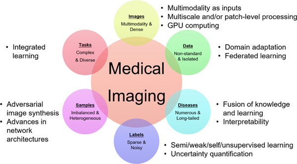
Since its renaissance, deep learning has been widely used in various medical imaging tasks and has achieved remarkable success in many medical imaging applications, thereby propelling us into the so-called artificial intelligence (AI) era. It is known that the success of AI is mostly attributed to the availability of big data with annotations for a single task and the advances in high performance computing. However, medical imaging presents unique challenges that confront deep learning approaches. In this survey paper, we first present traits of medical imaging, highlight both clinical needs and technical challenges in medical imaging, and describe how emerging trends in deep learning are addressing these issues. We cover the topics of network architecture, sparse and noisy labels, federating learning, interpretability, uncertainty quantification, etc. Then, we present several case studies that are commonly found in clinical practice, including digital pathology and chest, brain, cardiovascular, and abdominal imaging. Rather than presenting an exhaustive literature survey, we instead describe some prominent research highlights related to these case study applications. We conclude with a discussion and presentation of promising future directions.
Keywords: Medical imaging; deep learning; survey.
Figures
References
-
- Beutel J, Kundel HL, and Van Metter RL, Handbook of Medical Imaging. SPEI Press, 2000, vol. 1.
-
- Budovec JJ, Lam CA, and Kahn CE Jr, “Informatics in radiology: radiology gamuts ontology: differential diagnosis for the semantic web,” RadioGraphics, vol. 34, no. 1, pp. 254–264, 2014. - PubMed
-
- Rubin DL, “Informatics in radiology: measuring and improving quality in radiology: meeting the challenge with informatics,” Radiographics, vol. 31, no. 6, pp. 1511–1527, 2011. - PubMed
-
- Recht MP, Dewey M, Dreyer K, Langlotz C, Niessen W, Prainsack B, and Smith JJ, “Integrating artificial intelligence into the clinical practice of radiology: challenges and recommendations,” European Radiology, pp. 1–9, 2020. - PubMed
Grants and funding
- IK6 BX006185/BX/BLRD VA/United States
- U24 CA199374/CA/NCI NIH HHS/United States
- U01 CA239055/CA/NCI NIH HHS/United States
- R01 HL158071/HL/NHLBI NIH HHS/United States
- R01 CA268287/CA/NCI NIH HHS/United States
- R01 CA249992/CA/NCI NIH HHS/United States
- R01 HL151277/HL/NHLBI NIH HHS/United States
- R01 CA220581/CA/NCI NIH HHS/United States
- R01 CA216579/CA/NCI NIH HHS/United States
- R01 CA268207/CA/NCI NIH HHS/United States
- R01 CA202752/CA/NCI NIH HHS/United States
- R01 CA208236/CA/NCI NIH HHS/United States
- U01 CA248226/CA/NCI NIH HHS/United States
- I01 BX004121/BX/BLRD VA/United States
- R43 EB028736/EB/NIBIB NIH HHS/United States
- U01 CA269181/CA/NCI NIH HHS/United States
- R01 CA257612/CA/NCI NIH HHS/United States
- U54 CA254566/CA/NCI NIH HHS/United States
LinkOut - more resources
Full Text Sources
Other Literature Sources