Case Reports
. 2022 Nov 9;9(1):124-129.
doi: 10.1002/ibra.12074. eCollection 2023 Spring.

A typically progressive dissection of the internal carotid artery with recurrent hiccups: A case report with continuous 2-year data recording

Affiliations
Case Reports

A typically progressive dissection of the internal carotid artery with recurrent hiccups: A case report with continuous 2-year data recording

Zong-Min Zhang et al. Ibrain. .

Abstract

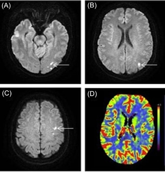
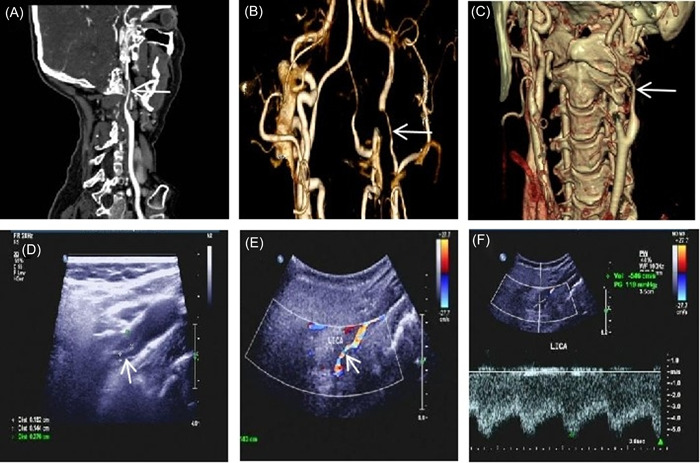
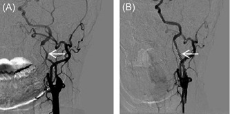
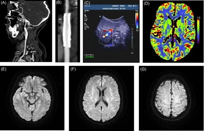
Patients with internal carotid artery dissection (ICAD) usually report headache, neck pain, Horner's syndrome, and ischemic stroke. Because the posterior cranial nerve is involved, some patients may show different forms of posterior cranial nerve paralysis. There have been no reports of patients with ICAD showing repeated hiccups. Here, to help clinicians identify ICAD early and gain a better understanding of the atypical manifestations of the disease, we report an atypical case of recurrent hiccup symptoms caused by ICAD.

Keywords: digital subtraction angiography (DSA); internal carotid artery dissection (ICAD); magnetic resonance imaging (MRI); recurrent hiccups.


Conflict of interest statement

The authors declare no conflict of interest.

Figures

Figure 1
Figure 1
(A–C) Brain diffusion‐weighted imaging showing multiple acute cerebral infarction in the left occipital lobe (white arrow) and parietal lobe. (D) Cerebral perfusion imaging showing decreased perfusion in the left cerebral hemisphere. [Color figure can be viewed at wileyonlinelibrary.com]
Figure 2
Figure 2
(A–C) Carotid computed tomography showing thin left internal carotid artery segment and critical stenosis of the lumen (white arrow). (D) Carotid ultrasound showing true and false double lumens in the left internal carotid artery, hypoechoic filling in the false lumen, and narrowing of the true lumen (white arrow); the residual inner diameter is about 1.4 mm and the original inner diameter was about 6.8 mm. (E) Color Doppler flow imaging examination showing no obvious blood flow signal in the false lumen, and a thin‐colored blood flow signal in the true lumen protruding outward (white arrow); the vascular diameter of the intermural hematoma is enlarged. (F) Pulsed wave measures the systolic flow rate of 546 cm/s at the stenosis of the lumen, and the distal blood flow spectrum shows low speed and low pulsation. [Color figure can be viewed at wileyonlinelibrary.com]
Figure 3
Figure 3
(A) On the left side, digital subtraction angiography shows the dissection from the cervical portion to the petrous portion, where a subtotal occlusion can be seen. The stenosis is about 45 mm long (white arrow). (B) The stent completely covers the stenosis and dissection opening (white arrow).
Figure 4
Figure 4
(A, B) Carotid artery computed tomography (CT) angiography. (C) Carotid artery ultrasound showing unobstructed blood flow in the stent. (D) Cranial CT perfusion imaging showing normal cerebral perfusion in both hemispheres. (E–G) Brain magnetic resonance imaging showing no infarction in the left occipital lobe and parietal lobe. [Color figure can be viewed at wileyonlinelibrary.com]
Figure 5
Figure 5
(A) The Xth cranial nerves is in close contact with the internal carotid artery at the level of the atlas. (B) The internal carotid artery is enlarged and stimulates the vagus nerve. [Color figure can be viewed at wileyonlinelibrary.com]

Similar articles

References

    1. Song JX, Lin XM, Hao ZQ, et al. Ocular manifestations of internal carotid artery dissection. Int J Ophthalmol. 2019;12(5):834‐839. 10.18240/ijo.2019.05.21 - DOI - PMC - PubMed
    1. Kurre W, Bansemir K, Aguilar Pérez M, et al. Endovascular treatment of acute internal carotid artery dissections: technical considerations, clinical and angiographic outcome. Neuroradiology. 2016;58(12):1167‐1179. 10.1007/s00234-016-1757-z - DOI - PubMed
    1. Fusco MR, Harrigan MR. Cerebrovascular dissections—a review part I: spontaneous dissections. Neurosurgery. 2011;68(1):242‐257, discussion 257. 10.1227/NEU.0b013e3182012323 - DOI - PubMed
    1. Nandwani K, Thng SY, Anaikatti P. An internal carotid artery dissection masquerading as optic neuritis: a case report. Cureus. 2021;13(1):e12810. 10.7759/cureus.12810 - DOI - PMC - PubMed
    1. Abdelmasih R, Abdelmaseih R, Monsour E, Reed J. Isolated third nerve palsy as a rare and solo presentation of internal carotid artery dissection in a young female—a surprising finding in the angiogram. Cureus. 2021;13(3):e14035. 10.7759/cureus.14035 - DOI - PMC - PubMed

Publication types