. 2023 Dec;101(12):1900-1913.
doi: 10.1002/jnr.25249. Epub 2023 Oct 3.

Breast-to-brain metastasis is exacerbated with chemotherapy through blood-cerebrospinal fluid barrier and induces Alzheimer's-like pathology

Affiliations

Breast-to-brain metastasis is exacerbated with chemotherapy through blood-cerebrospinal fluid barrier and induces Alzheimer's-like pathology

B Saatian et al. J Neurosci Res. 2023 Dec.

Abstract

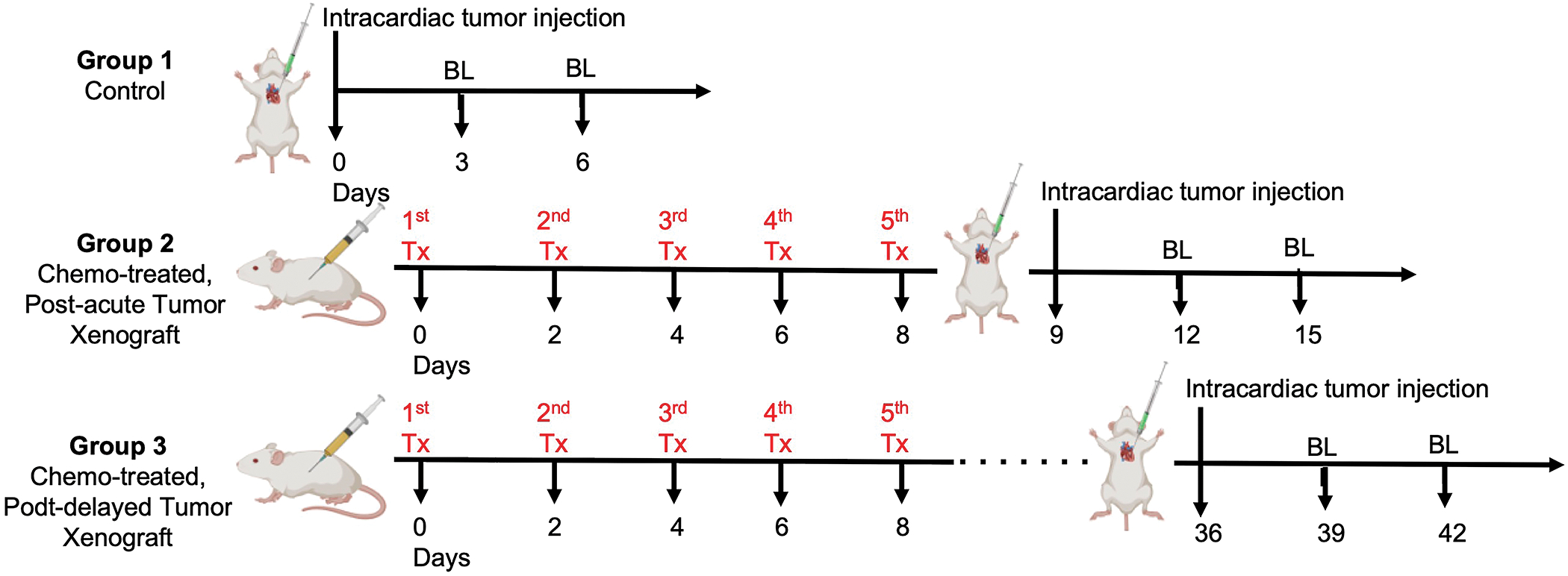
Control of breast-to-brain metastasis remains an urgent unmet clinical need. While chemotherapies are essential in reducing systemic tumor burden, they have been shown to promote non-brain metastatic invasiveness and drug-driven neurocognitive deficits through the formation of neurofibrillary tangles (NFT), independently. Now, in this study, we investigated the effect of chemotherapy on brain metastatic progression and promoting tumor-mediated NFT. Results show chemotherapies increase brain-barrier permeability and facilitate enhanced tumor infiltration, particularly through the blood-cerebrospinal fluid barrier (BCSFB). This is attributed to increased expression of matrix metalloproteinase 9 (MMP9) which, in turn, mediates loss of Claudin-6 within the choroid plexus cells of the BCSFB. Importantly, increased MMP9 activity in the choroid epithelium following chemotherapy results in cleavage and release of Tau from breast cancer cells. This cleaved Tau forms tumor-derived NFT that further destabilize the BCSFB. Our results underline for the first time the importance of the BCSFB as a vulnerable point of entry for brain-seeking tumor cells post-chemotherapy and indicate that tumor cells themselves contribute to Alzheimer's-like tauopathy.

Keywords: CSF; blood-cerebrospinal fluid barrier; brain metastasis; breast cancer; chemotherapy; tau.


Conflict of interest statement

Conflict of Interest Statement

Authors declare that they have no conflicting interests.

Figures

Fig 1.
Fig 1.. Chemotherapy-induced brain metastasis model.
Study design to determine the effect of brain permeability for control (Group 1), acute (Group 2) and delayed (Group 3) adjuvant chemotherapy on initial brain colonization from systemic tumor cells.
Fig. 2.
Fig. 2.. Chemotherapy facilitates breast cancer cells’ entry into the brain through the BCSFB.
In vivo analysis of brain permeability via Fluorescein-dye uptake in the brain of mice treated with paclitaxel (A) and 5-FU (B) compared to vehicle group. (C) In vivo BLI and (D) quantification of BBM signal in control (Group I), acute chemo-treated (Group II), and delayed chemo-treated (Group III) groups 3 days and 6 days post tumor injection (DPTI). Quantification (E) and imaging (F) of metastatic tumor-GFP fluorescence intensity in the brain parenchyma (blue) and in the choroid plexus of the BCSFB of the lateral and 4th ventricles (red) in Groups 1–3. (G) In vitro migration capacity of BC cells across the BBB versus BCSFB in control and paclitaxel-treatments. (H) MRI and quantification (I) of patients with brain metastasis having peri-atrial brain metastasis (red) with posterior CP blood supply involvement; Anterior of lateral ventricle (A of LV). (J) Representative histological H&E sections from whole-brain patient tissue from rapid autopsy cases with parenchymal BMs and tumor cells in the ipsilateral BCSFB. (K) Quantification of extravascular extracellular space (Ve) in the choroid plexus from DCE-MRI of patients with and without chemotherapy exposure prior to brain metastasis diagnosis. (L-M) Representative examples of Ve color map from DCE-MRI from both groups shown in the left glomus of the CP.
Fig. 3.
Fig. 3.. Chemotherapy upregulates CP-MMP9 leading to downregulation of CLDN6 tight junctions on BCSFB.
(A) In vitro BCSFB permeability assessed by fluorescein-dye on naïve CP (control), paclitaxel (Pac)-treated CP cells, or CP cells exposed to MDA-MB-231 condition media (CM) alone or MDA-MB-231CM pre-treated with paclitaxel. (B) Clustergram representing mRNA expression of junctional markers in CP cells treated with paclitaxel (PTX), MDA-MB-231 CM, MDA-MB-231 CM pre-treated with PTX, and co-cultured with MDA-MB-231 alone or MDA-MB-231 pretreated with PTX. Color scheme goes from red for no change in expression to purple for down-regulated genes. (C) Venn diagram of downregulated junctional markers in all five conditions from (B) relative to control. (D) RT-qPCR validation of four common junctional markers downregulated in all five tested conditions in CP cells. (E) Immunofluorescent (IF) imaging of Claudin-6 in choroid plexus adjacent to tumor lesion compared to CP distal to metastatic lesion in post-mortem patient tissue diagnosed with brain metastasis. Images taken at 40X. (F) IF imaging of MMP9 in CP of the BCSFB ipsilateral to tumor lesion compared to choroid plexus on the contralateral hemisphere in post-mortem patient tissue diagnosed with brain metastasis. Images taken at 40X. (G) MMP9 expression in CP cells treated with paclitaxel, tumor condition media (CM), and combination of tumor condition media and paclitaxel relative to CP cells alone in vitro. (H) Quantification of exogenous MMP9 released by CP treated alone, with primary breast cancer MDA-MB-231, or BBM3.1 CM ± paclitaxel in vitro. (I) RT-qPCR analysis of junctional markers’ expression in CP KDMMP9 cells treated with tumor CM ± paclitaxel. (J) IF imaging of Claudin-6 in CP KDMMP9 cells treated with tumor CM ± paclitaxel. Images taken at 63X (K) Flourescein dye-assay determining permeability of BCSFB (control CP and CP KDMMP9) when treated with breast cancer cells (MDA-MB-231 CM ± paclitaxel).
Fig. 4.
Fig. 4.. Tumors cells highjack CP-MMP9 leading to NFT and subsequent increase in BCSF permeability.
(A) mRNA analysis of MAPT 3R and MAPT 4R expression and their (B) ratio in primary breast cancer MDA-MB-231 and BT-474, and USC-BBM3.1. (C) qPCR validation of total MAPT, MAPT 3R/4R knockdown in MDA-MB-231 cells (D) BLI of MDA-MB-231 and MDA-MB-231 KDMAPT xenografts in vivo. Kaplan-Meier survival analyses of breast cancer MDA-MB-231-bearing and MDA-MB-231 KDMAPT mice. (E) Quantification of Tau NFT formation by PHF staining in BC cells treated with DMSO and Paclitaxel. (F) Expression of junctional markers in BCSFB treated with exogenous tau, and CM from MDA-MB-231 and MDA-MB-231 KDMAPT. (G) Quantification of BCSFB permeability in vitro in CP cells treated with CM from MDA-MB-231 and MDA-MB-231 KDMAPT using fluorescein assay. (H) Quantification of exogenous MMP9 released from CP alone, treated with CM from MDA-MB-231 (Control), or MDA-MB-231 KDMAPT, and from MDA-MB-231 or MDA-MB-231 KDMAPT. (I) PHF quantification in the condition media of CP cells alone, CP KDMMP9 cells, CP cells treated with CM from MDA-MB-231, CP KDMMP9 cells treated with CM from MDA-MB-231, and in MD-MB-231 CM. (J) In vitro migration assay through BCSFB quantifying the migrational capacity of MDA-MB-231 and MDA-MB-231 KDMAPT cells across the wild-type CP (Control) and CP KDMMP9 cells. (K) MAPT expression in cells found in CSF of a leptomeningeal (LMD) patient using single cell RNA-Seq analysis. (L) Tau quantification in the CSF of patients ± LMD. (M) PHF quantification in of CP cells (control, CP KDMMP9) treated with CM from MDA-MB-231 cells treated with Paclitaxel. (N) PHF quantification of CP cells treated with CM from MDA-MB-231 KDMAPT cells in two conditions (cells only, cells treated with Paclitaxel) (O) Accumulation of corpora amylacea waste vacuoles on ipsilateral brain parenchyma to tumor lesion, adjacent to lateral ventricle (LV) CSF, and CP in H&E sections from post-mortem brain metastatic patient tissue. Images taken at 40X.

References

    1. Abbott NJ, Patabendige AA, Dolman DE, Yusof SR, & Begley DJ (2010). Structure and function of the blood-brain barrier. Neurobiol Dis, 37(1), 13–25. doi:10.1016/j.nbd.2009.07.030 - DOI - PubMed
    1. Abu Samaan TM, Samec M, Liskova A, Kubatka P, & Büsselberg D (2019). Paclitaxel’s Mechanistic and Clinical Effects on Breast Cancer. Biomolecules, 9(12). doi:10.3390/biom9120789 - DOI - PMC - PubMed
    1. Ahles TA, Root JC, & Ryan EL (2012). Cancer- and cancer treatment-associated cognitive change: an update on the state of the science. Journal of clinical oncology : official journal of the American Society of Clinical Oncology, 30(30), 3675–3686. doi:10.1200/JCO.2012.43.0116 - DOI - PMC - PubMed
    1. Barghorn S, Davies P, & Mandelkow E (2004). Tau paired helical filaments from Alzheimer’s disease brain and assembled in vitro are based on beta-structure in the core domain. Biochemistry, 43(6), 1694–1703. doi:10.1021/bi0357006 - DOI - PubMed
    1. Brezden CB, Phillips KA, Abdolell M, Bunston T, & Tannock IF (2000). Cognitive function in breast cancer patients receiving adjuvant chemotherapy. Journal of clinical oncology : official journal of the American Society of Clinical Oncology, 18(14), 2695–2701. doi:10.1200/JCO.2000.18.14.2695 - DOI - PubMed

Publication types

Substances

LinkOut - more resources