Review
. 2023 Aug;248(15):1333-1346.
doi: 10.1177/15353702231191186. Epub 2023 Oct 3.

Ubiquitination detection techniques

Affiliations
Review

Ubiquitination detection techniques

Anthony Ruvindi I De Silva et al. Exp Biol Med (Maywood). 2023 Aug.

Abstract

Ubiquitination is an intricately regulated post-translational modification that involves the covalent attachment of ubiquitin to a substrate protein. The complex dynamic nature of the ubiquitination process regulates diverse cellular functions including targeting proteins for degradation, cell cycle, deoxyribonucleic acid (DNA) damage repair, and numerous cell signaling pathways. Ubiquitination also serves as a crucial mechanism in protein quality control. Dysregulation in ubiquitination could result in lethal disease conditions such as cancers and neurodegenerative diseases. Therefore, the ubiquitination cascade has become an attractive target for therapeutic interventions. Enormous efforts have been made to detect ubiquitination involving different detection techniques to better grasp the underlying molecular mechanisms of ubiquitination. This review discusses a wide range of techniques stretching from the simplest assays to real-time assays. This includes western blotting/immunoblotting, fluorescence assays, chemiluminescence assays, spectrophotometric assays, and nanopore sensing assays. This review compares these applications, and the inherent advantages and limitations.

Keywords: Ubiquitination; chemiluminescence; detection techniques; fluorescence; spectrophotometric.


Conflict of interest statement

Declaration Of Conflicting InterestsThe author(s) declared no potential conflicts of interest with respect to the research, authorship, and/or publication of this article.

Figures

Figure 1.
Figure 1.
Overview of the UbiReal approach: (A) Schematic representation of the ubiquitin conjugation steps monitored through the UbiReal approach. (B) Data representing the UbiReal approach monitoring the ubiquitin conjugation. A fluorescently labeled ubiquitin was used to track the progression through the cascade involving UBE1 (E1 enzyme), UB2D3 (E2 enzyme), and NleL (E3 enzyme) with significant molecular weights. A large FP signal was produced in the activation step for the conjugation of ubiquitin with UBE with a large molecular weight. Then, the conjugation step involving UBE2D3 with an intermediate molecular weight resulted in a corresponding FP signal. Next, the ligation step involving NleL caused an increase in the FP signal over time due to the addition of unlabeled ubiquitin into the solution, forming a polyubiquitin chain. FP: Fluorescence polarization. Source: Reprinted from Franklin and Pruneda. Copyright (2019), with permission from Frontiers.
Figure 2.
Figure 2.
A schematic representation of the overview of fluorescent confocal on-bead assay utilizing ubiquitin-activating enzyme (UBE1), ubiquitin conjugating enzyme (UBE2 L3), E6AP (ubiquitin-ligating enzyme), fluorescently labeled ubiquitin using Cy5 dye (Cy5-ubiquitin), and ATP. Initially, E6AP was immobilized on nickel nitrilotriacetic acid (Ni2 + nitrilotriacetic acid) agarose microbeads and distributed in a microwell plate. Then, the reaction mixture was added, and the reactions were monitored over time. The ubiquitination reaction was monitored by detecting the enzymatic conjugation of labeled ubiquitin onto microbeads, through confocal imaging using an Opera™ High Content Screening System. The conjugation caused the formation of a fluorescent “ring” in the image plane across the microbeads, and the intensity of the fluorescence emission was determined over time. ATP: adenosine triphosphate. Source: Reprinted from Koszela et al. Copyright (2018), with permission from Springer Nature.
Figure 3.
Figure 3.
A schematic representation of the molybdenum blue assay. The adenylation of ubiquitin by E1 released pyrophosphate product during the formation of ubiquitin AMP which was then converted to two phosphates via pyrophosphatase. The progression of the reaction was halted, and the phosphomolybdate formation was initiated by the addition of ascorbic acid and ammonium molybdate. Next, the color of the solution was developed using citric acid and sodium arsenite, and the absorbance of phosphomolybdate was measured at 850 nm. Since two phosphate molecules were produced for every ubiquitin transferred, the absorbance was halved and corrected for the background ATP hydrolysis. At the end, the concentration of the phosphate was determined from the corrected absorbance by comparing it to a standard curve. ATP: adenosine triphosphate. Source: Reprinted from Berndsen and Wolberger. Copyright (2011), with permission from Elsevier.
Figure 4.
Figure 4.
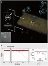
A schematic representation of the passage of di-ubiquitin molecules through the nanopore. The inset for the transmission electron microscopy image displays the solid-state nanopores of 3.5 nm used as single-molecule sensors for ubiquitin and ubiquitin chains. Source: Reprinted from Nir et al. Copyright (2015), with permission from Elsevier.

References

    1. Swatek KN, Komander D. Ubiquitin modifications. Cell Res 2016;26:399–422 - PMC - PubMed
    1. Sun L, Chen ZJ. The novel functions of ubiquitination in signaling. Curr Opin Cell Biol 2004;16:119–26 - PubMed
    1. Popovic D, Vucic D, Dikic I. Ubiquitination in disease pathogenesis and treatment. Nat Med 2014;20:1242–53 - PubMed
    1. Finley D, Ciechanover A, Varshavsky A. Ubiquitin as a central cellular regulator. Cell 2004;116:S29–32 - PubMed
    1. Haas AL, Rose IA. The mechanism of ubiquitin activating enzyme. A kinetic and equilibrium analysis. J Biol Chem 1982;257:10329–37 - PubMed

Publication types

MeSH terms