. 2023 Dec;12(6):3263-3279.
doi: 10.1007/s40123-023-00815-9. Epub 2023 Oct 3.

Repeatability and Interobserver Reproducibility of a Swept-Source Optical Coherence Tomography for Measurements of Anterior, Posterior, and Total Corneal Power

Affiliations

Repeatability and Interobserver Reproducibility of a Swept-Source Optical Coherence Tomography for Measurements of Anterior, Posterior, and Total Corneal Power

Chak Seng Lei et al. Ophthalmol Ther. 2023 Dec.

Abstract

Introduction: The aim of this work is to evaluate the intraobserver repeatability and interobserver reproducibility of corneal power measurements obtained with a swept-source optical coherence tomographer (CASIA 2, Tomey, Japan) in healthy subjects.

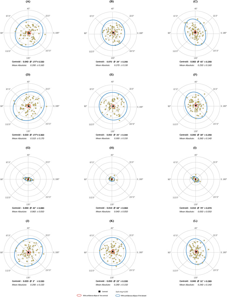
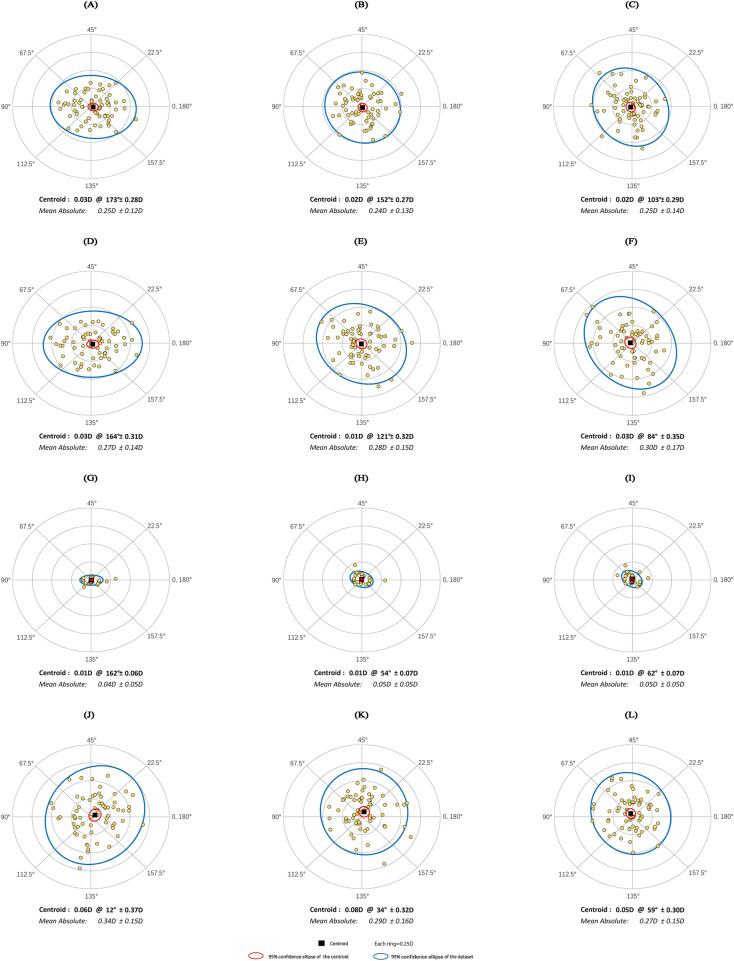
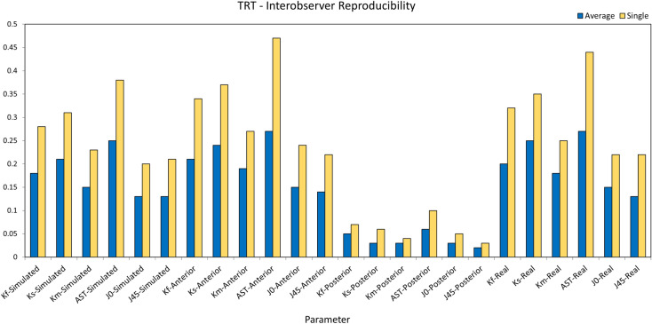
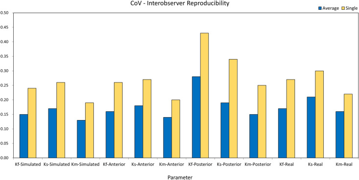
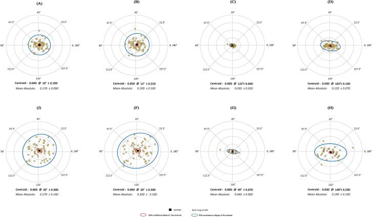
Methods: A total of 67 right eyes from 67 healthy subjects were enrolled. Two experienced observers measured each eye three times consecutively with the CASIA 2. Corneal power values were recorded as simulated keratometry, anterior, posterior, and total corneal power. Parameters were flattest keratometry (Kf), steepest keratometry (Ks), mean keratometry (Km), astigmatism magnitude, astigmatism power vectors J0 and J45. Intraobserver repeatability and interobserver reproducibility of the CASIA 2 were assessed by the within-subject standard deviation (Sw), test-retest repeatability (TRT), coefficients of variation (CoV), and intraclass correlation coefficients (ICCs). Double-angle plots were used for astigmatism vector analysis.

Results: The CASIA 2 had high repeatability for all corneal power values, with Sw values ≤ 0.17 diopters (D), TRT ≤ 0.46 D, and ICCs ranging from 0.866 to 0.998. Interobserver reproducibility was also high, showing all Sw values ≤ 0.10 D, TRT ≤ 0.27 D, and ICCs ≥ 0.944. The reproducibility of the average of three consecutive measurements (Sw 0.01-0.10 D, TRT 0.03-0.27 D, ICC 0.944-0.998) was higher than the reproducibility of single measurements (Sw 0.01-0.17 D, TRT 0.03-0.47 D, ICC 0.867-0.996).

Conclusions: The CASIA 2 showed high intraobserver repeatability and interobserver reproducibility for anterior, posterior, and total corneal power measurements in 6.0-mm diameter area. In addition, we suggest that using the average of three consecutive measurements can improve reproducibility between observers, compared to single measurements only.

Keywords: Biometry; Corneal power; Swept-source optical coherence tomographer.


Conflict of interest statement

Chak Seng Lei, Xuanqiao Lin, Rui Ning, Jinjin Yu, Xiaomin Huang, Kexin Li, Yiran Wang, Giacomo Savini, Domenico Schiano-Lomoriello, Xingtao Zhou, and Jinhai Huang have nothing to disclose.

Figures

Fig. 1
Fig. 1
Double-angle plots showing intraobserver differences in astigmatism between the first observer’s repeated measurements in each corneal power measurement. Each ring represents 0.25 diopters (D), and the outer ring represents 1.0 D. The small ellipses (red) show the 95% confidence ellipses for the centroid and the larger ellipses (blue) show the 95% confidence ellipse for the difference vectors of the dataset. First row: Simulated keratometry. Second row: Anterior corneal power. Third row: Posterior corneal power. Fourth row: Total corneal power. First column: difference between first and second measurements. Second column: difference between first and third measurements. Third column: difference between second and third measurements
Fig. 2
Fig. 2
Double-angle plots showing intraobserver differences in astigmatism between the second observer’s repeated measurements in each corneal power measurement. Each ring represents 0.25 diopters (D), and the outer ring represents 1.0 D. The small ellipses (red) show the 95% confidence ellipses for the centroid and the larger ellipses (blue) show the 95% confidence ellipse for the difference vectors of the dataset. First row: Simulated keratometry. Second row: Anterior corneal power. Third row: Posterior corneal power. Fourth row: Total corneal power. First column: difference between first and second measurements. Second column: difference between first and third measurements. Third column: difference between second and third measurements
Fig. 3
Fig. 3
Test–retest repeatability (TRT) values of all measured parameters for averaged measurements and single measurement. Kf flattest keratometry, Ks steepest keratometry, Km mean keratometry, AST astigmatism magnitude, J0 cylinder axis at the 180° and 90° meridians, J45 cylinder axis at the 45° and 135° meridians
Fig. 4
Fig. 4
Coefficients of variation (CoV) values of keratometric parameters for averaged measurements and single measurement. Kf flattest keratometry, Ks steepest keratometry, Km mean keratometry
Fig. 5
Fig. 5
Double-angle plots showing interobserver differences in astigmatism in each corneal power measurement. Each ring represents 0.25 diopters (D), and the outer ring represents 1.0 D. The small ellipses (red) show the 95% confidence ellipses for the centroid and the larger ellipses (blue) show the 95% confidence ellipse for the difference vectors of the dataset. First row: Average method. Second row: Single method. First column: Simulated keratometry. Second column: Anterior corneal power. Third column: Posterior corneal power. Fourth column: Total corneal power

References

    1. Tomidokoro A, Oshika T, Amano S, Higaki S, Maeda N, Miyata K. Changes in anterior and posterior corneal curvatures in keratoconus. Ophthalmology. 2000;107(7):1328–1332. doi: 10.1016/S0161-6420(00)00159-7. - DOI - PubMed
    1. Olsen T. Calculation of intraocular lens power: a review. Acta Ophthalmol Scand. 2007;85(5):472–485. doi: 10.1111/j.1600-0420.2007.00879.x. - DOI - PubMed
    1. Hirnschall N, Findl O, Bayer N, et al. Sources of error in toric intraocular lens power calculation. J Refract Surg Thorofare NJ 1995. 2020;36(10):646–652. doi: 10.3928/1081597X-20200729-03. - DOI - PubMed
    1. Koch DD, Ali SF, Weikert MP, Shirayama M, Jenkins R, Wang L. Contribution of posterior corneal astigmatism to total corneal astigmatism. J Cataract Refract Surg. 2012;38(12):2080–2087. doi: 10.1016/j.jcrs.2012.08.036. - DOI - PubMed
    1. Gutmark R, Guyton DL. Origins of the keratometer and its evolving role in ophthalmology. Surv Ophthalmol. 2010;55(5):481–497. doi: 10.1016/j.survophthal.2010.03.001. - DOI - PubMed

LinkOut - more resources