. 2023 Oct 3;19(10):e1011662.
doi: 10.1371/journal.ppat.1011662. eCollection 2023 Oct.

VP2 residue N142 of coxsackievirus A10 is critical for the interaction with KREMEN1 receptor and neutralizing antibodies and the pathogenicity in mice

Affiliations

VP2 residue N142 of coxsackievirus A10 is critical for the interaction with KREMEN1 receptor and neutralizing antibodies and the pathogenicity in mice

Xue Li et al. PLoS Pathog. .

Abstract

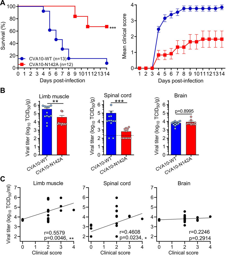
Coxsackievirus A10 (CVA10) has recently emerged as one of the major causative agents of hand, foot, and mouth disease. CVA10 may also cause a variety of complications. No approved vaccine or drug is currently available for CVA10. The residues of CVA10 critical for viral attachment, infectivity and in vivo pathogenicity have not been identified by experiment. Here, we report the identification of CVA10 residues important for binding to cellular receptor KREMEN1. We identified VP2 N142 as a key receptor-binding residue by screening of CVA10 mutants resistant to neutralization by soluble KREMEN1 protein. The receptor-binding residue N142 is exposed on the canyon rim but highly conserved in all naturally occurring CVA10 strains, which provides a counterexample to the canyon hypothesis. Residue N142 when mutated drastically reduced receptor-binding activity, resulting in decreased viral attachment and infection in cell culture. More importantly, residue N142 when mutated reduced viral replication in limb muscle and spinal cord of infected mice, leading to lower mortality and less severe clinical symptoms. Additionally, residue N142 when mutated could decrease viral binding affinity to anti-CVA10 polyclonal antibodies and a neutralizing monoclonal antibody and render CVA10 resistant to neutralization by the anti-CVA10 antibodies. Overall, our study highlights the essential role of VP2 residue N142 of CVA10 in the interactions with KREMEN1 receptor and neutralizing antibodies and viral virulence in mice, facilitating the understanding of the molecular mechanisms of CVA10 infection and immunity. Our study also provides important information for rational development of antibody-based treatment and vaccines against CVA10 infection.


Conflict of interest statement

The authors have declared that no competing interests exist.

Figures

Fig 1
Fig 1. Selection of CVA10 mutants resistant to neutralization with soluble human KRM1 receptor.
(A) A flowchart of the screening procedure. (B) Screening and information of soluble KRM1-resistant mutants. Green circle, no CPE; orange, partial CPE; red, complete CPE. 10 and 9 plaques were isolated from well #1 (the upper well) and #2 (the lower well), respectively, and then sequenced to identify mutations. Viral capsid residues are numbered from 1001, 2001, and 3001 in VP1, VP2, and VP3, respectively. (C) A summary of the three major types of the mutations. The red arrows indicate the substitutions in the first or second nucleotides of the codons.
Fig 2
Fig 2. The role of the mutations in conferring resistance to neutralization with soluble KRM1-Fc.
(A) Wildtype and mutant CVA10 viruses were titrated by TCID50 assay and analyzed for resistance to neutralization with KRM1-Fc. Fold increase or decrease (prefixed with a minus sign) in viral titer and neutralization concentration of KRM1-Fc against the mutants, relative to wildtype CVA10. Red highlighting, more than 10-fold decrease. (B) Schematic representation of construction of the T7 promoter (PT7)-driven infectious clone of CVA10. The indicated mutations were separately introduced into the infectious clone. (C) Schematic of rescue and characterization of wildtype and mutant CVA10 viruses. T7 RNA-pol, T7 RNA polymerase. (D) The rescued CVA10 viruses were titrated by TCID50 assay and sequenced. dpi, day post infection. (E) The rescued CVA10 viruses were analyzed for resistance to neutralization with KRM1-Fc.
Fig 3
Fig 3. Location and conservation of the mutated residues.
(A) Surface representation of the structure of CVA10 alone and in complex with KRM1 receptor (PDB: 7BZU). Upper left panel: the structure of one asymmetric unit of CVA10. VP1, light blue; VP2, dark sea green; VP3, light coral. VP1’, the C terminus of an adjacent VP1 is colored in light steel blue. Upper right panel: the canyon topology. 5, 3, and 2: five-, three-, and two-fold axis. Lower left panel: the structure of one asymmetric unit of CVA10 in complex with KRM1. KRM1, medium violet red. Lower right panel: KRM1-contacting residues of CVA10 (distance cut-off: 4 Å) are colored in medium violet red. (B) The detailed interactions between viral residues (N2142, N3183, and T1219) and surrounding KRM1 residues. Black dash lines indicate hydrogen bonds. (C) The impact of mutation of N2142 on the virus-receptor interaction was analyzed using SWISS-MODEL. Nitrogen atom, blue; oxygen, red; carbon, gray. (D) Sequence conservation analysis of CVA10 residues 2142, 3183, and 1219. CVA10 VP2, VP3, VP1 protein sequences were obtained from NCBI database and used for this analysis.
Fig 4
Fig 4. N2142A mutation reduced viral replication and binding to cell by impairing receptor binding.
(A) Growth kinetics of rescued CVA10-WT and CVA10-N142A were compared in RD cells. RD cells were infected at a MOI of 0.01 and viral titers were determined at the indicated time points post infection. Data are means ± SD of three replicate samples. Analysis was performed using two-way ANOVA. ns, no significant difference (p ≥ 0.05); **, p < 0.01; ****, p < 0.0001. (B) CVA10-WT or CVA10-N142A infected cell cultures were subjected to sucrose gradient ultracentrifugation, and the resulting 12 fractions were analyzed by SDS-PAGE. The fraction #10 samples (indicated by red arrows) were selected for subsequent analysis. (C) The fraction #10 samples of CVA10-WT and CVA10-N142A were diluted to 50 μg/ml and subjected to negative stain electron microscopy. Red and blue arrows indicate full (mature virion) and empty CVA10 particles, respectively. (D) The fraction #10 samples of CVA10-WT and CVA10-N142A were diluted to 1 μg/ml and assayed for viral titers. Fold decrease (prefixed with a minus sign) in viral titer is also shown. (E) 100 ng/ml of CVA10 viral particles (fraction #10) were attached to prechilled RD cells at 4°C for 1 h, and after washing, the levels of CVA10 RNA were determined by RT-qPCR. Data are means ± SD of four replicate samples. The Mann–Whitney test was used to compare differences. *, p < 0.05. (F) Reactivity of CVA10-WT and CVA10-N142A viral particles with human KRM1-Fc protein were determined by ELISA. CVA10 viral particles were serially diluted and coated onto microplates, followed by incubation with biotinylated KRM1-Fc or ACE2-Fc (ctr) protein and HRP-conjugated streptavidin. Data are means ± SD of three replicate samples. (G) Proposed model of residue N2142-dependent CVA10 attachment and infection of target cells. The N2142A mutation is indicated by red dots.
Fig 5
Fig 5. N2142A mutation could reduce mortality rate and severity of symptoms by decreasing viral loads in limb muscle and spinal cord.
(A) Groups of 2-day-old ICR mice were inoculated with 300 TCID50 of CVA10-WT or CVA10-N142A, and then survival (left panel) and clinical symptoms (right panel) were observed daily for 14 days. Clinical scores were graded as follows: 0, healthy; 1, reduced mobility; 2, limb weakness; 3, limb paralysis; 4, death. Number of mice per group were indicated in brackets. Statistical significance of survival curves between groups was determined by Log-rank (Mantel-Cox) test. ***, p < 0.001. All error bars represent SEM. (B) Groups of ICR mice infected with CVA10-WT or CVA10-N142A were sacrificed at 4 dpi, and limb muscle (left panel), spinal cord (middle panel), and brain (right panel) were collected, weighed, and tested for virus titers. There were 12 mice per group. Each symbol represents an individual mouse. The Mann–Whitney test was used to compare differences. **, p < 0.01; ***, p < 0.001. (C) Correlations between viral titers in the indicated organs and clinical scores. Viral titers and clinical scores of individual infected mice from the CVA10-WT and CVA10-N142A groups were used for Pearson correlation coefficient analysis. *, p < 0.05; **, p < 0.01.
Fig 6
Fig 6. N2142A mutation could lead to milder limb muscle pathology in infected mice and significantly reduce CVA10 binding affinity to murine KRM1 receptor.
(A) Groups of ICR mice inoculated with medium (control), CVA10-WT, or CVA10-N142A were sacrificed at 4 dpi, and limb muscle samples were collected for histological examination. Histopathologic scores were graded according to the severity of myositis as: not present, mild, moderate, and severe. Number of mice (n) is shown. (B) Histopathologic scores of the CVA10-WT and CVA10-N142A groups. Each symbol represents an individual mouse. The Mann–Whitney test was used to compare differences. **, p < 0.01. The solid line indicates the mean values. (C) Correlations between histopathologic scores and clinical scores. Histopathologic scores of individual mice from the CVA10-WT and CVA10-N142A groups were used for Pearson correlation analysis. *, p < 0.05. (D) Reactivity of CVA10-WT and CVA10-N142A viral particles with murine KRM1-Fc (mKrm1-Fc) were analyzed by ELISA. Serially diluted Viral particles were coated onto microplates and then incubated with biotinylated KRM1-Fc or ACE2-Fc (ctr) protein and HRP-conjugated streptavidin. Data are means ± SD of three replicate samples. (E) Sequence alignment of the human (hKRM1) and mouse (mKrm1) KRM1 proteins. The three KRM1 residues (D88, G89, and D90) predicted to contact CVA10 residue N142 are boxed. The graph was generated using CLC Sequence Viewer software.
Fig 7
Fig 7. CVA10-N142A is more resistant to anti-CVA10 sera and MAb 2A11 as compared to CVA10-WT.
(A-B) The reactivities of anti-CVA10 sera (A) and MAb 2A11 (B) to CVA10-WT and CVA10-N142A were determined by ELISA. Purified viral particles were serially diluted and coated onto ELISA plates. Data are mean ± SD of triplicate wells. (C-D) Neutralization activities of anti-CVA10 sera (C) and MAb 2A11 (D) against CVA10-WT and CVA10-N142A were determined. CVA10 viruses were incubated with two-fold serial dilutions of anti-CVA10 antibodies for 1 h before adding to RD cells. Cell viability was measured 3 days after culture. The red dashed line indicates NT50 or IC50. Data are mean ± SEM of four replicate wells.

Similar articles

Cited by

References

    1. Bian L, Gao F, Mao Q, Sun S, Wu X, Liu S, et al.. Hand, foot, and mouth disease associated with coxsackievirus A10: more serious than it seems. Expert Rev Anti Infect Ther. 2019;17(4):233–42. Epub 2019/02/23. doi: 10.1080/14787210.2019.1585242 . - DOI - PubMed
    1. Banno F, Shibata S, Hasegawa M, Matsuoka S, Okumura A. Acute flaccid myelitis presumably caused by coxsackie virus A10. Pediatr Int. 2021;63(1):104–5. Epub 2021/01/16. doi: 10.1111/ped.14361 . - DOI - PubMed
    1. Lu QB, Zhang XA, Wo Y, Xu HM, Li XJ, Wang XJ, et al.. Circulation of Coxsackievirus A10 and A6 in hand-foot-mouth disease in China, 2009–2011. PLoS One. 2012;7(12):e52073. Epub 2012/12/29. doi: 10.1371/journal.pone.0052073 ; PubMed Central PMCID: PMC3525556. - DOI - PMC - PubMed
    1. Fuschino ME, Lamson DM, Rush K, Carbone LS, Taff ML, Hua Z, et al.. Detection of coxsackievirus A10 in multiple tissues of a fatal infant sepsis case. J Clin Virol. 2012;53(3):259–61. Epub 2012/01/03. doi: 10.1016/j.jcv.2011.12.011 S1386-6532(11)00518-X [pii]. . - DOI - PubMed
    1. Ji T, Guo Y, Huang W, Shi Y, Xu Y, Tong W, et al.. The emerging sub-genotype C2 of CoxsackievirusA10 Associated with Hand, Foot and Mouth Disease extensively circulating in mainland of China. Sci Rep. 2018;8(1):13357. Epub 2018/09/08. doi: 10.1038/s41598-018-31616-x [pii]. ; PubMed Central PMCID: PMC6127217. - DOI - PMC - PubMed

Publication types

MeSH terms

LinkOut - more resources