Molecular View on the i RGD Peptide Binding Mechanism: Implications for Integrin Activity and Selectivity Profiles
- PMID: 37788340
- PMCID: PMC10598797
- DOI: 10.1021/acs.jcim.3c01071
Molecular View on the i RGD Peptide Binding Mechanism: Implications for Integrin Activity and Selectivity Profiles
Abstract
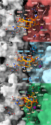
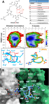
Receptor-selective peptides are widely used as smart carriers for specific tumor-targeted delivery. A remarkable example is the cyclic nonapeptide iRGD (CRGDKPGDC, 1) that couples intrinsic cytotoxic effects with striking tumor-homing properties. These peculiar features are based on a rather complex multistep mechanism of action, where the primary event is the recognition of RGD integrins. Despite the high number of preclinical studies and the recent success of a phase I trial for the treatment of pancreatic ductal adenocarcinoma (PDAC), there is little information available about the iRGD three-dimensional (3D) structure and integrin binding properties. Here, we re-evaluate the peptide's affinity for cancer-related integrins including not only the previously known targets αvβ3 and αvβ5 but also the αvβ6 isoform, which is known to drive cell growth, migration, and invasion in many malignancies including PDAC. Furthermore, we use parallel tempering in the well-tempered ensemble (PT-WTE) metadynamics simulations to characterize the in-solution conformation of iRGD and extensive molecular dynamics calculations to fully investigate its binding mechanism to integrin partners. Finally, we provide clues for fine-tuning the peptide's potency and selectivity profile, which, in turn, may further improve its tumor-homing properties.
Conflict of interest statement
The authors declare no competing financial interest.
Figures


Similar articles
-
Integrin-targeted delivery into cancer cells of a Pt(IV) pro-drug through conjugation to RGD-containing peptides.Dalton Trans. 2015 Jan 7;44(1):202-12. doi: 10.1039/c4dt02710h. Dalton Trans. 2015. PMID: 25369773
-
Targeted Modification of the Cationic Anticancer Peptide HPRP-A1 with iRGD To Improve Specificity, Penetration, and Tumor-Tissue Accumulation.Mol Pharm. 2019 Feb 4;16(2):561-572. doi: 10.1021/acs.molpharmaceut.8b00854. Epub 2019 Jan 14. Mol Pharm. 2019. PMID: 30592418
-
Distinct structural requirements for binding of the integrins alphavbeta6, alphavbeta3, alphavbeta5, alpha5beta1 and alpha9beta1 to osteopontin.Matrix Biol. 2005 Sep;24(6):418-27. doi: 10.1016/j.matbio.2005.05.005. Matrix Biol. 2005. PMID: 16005200
-
iRGD: A Promising Peptide for Cancer Imaging and a Potential Therapeutic Agent for Various Cancers.J Oncol. 2019 Jun 26;2019:9367845. doi: 10.1155/2019/9367845. eCollection 2019. J Oncol. 2019. PMID: 31346334 Free PMC article. Review.
-
Radiolabeled Cyclic RGD Peptide Bioconjugates as Radiotracers Targeting Multiple Integrins.Bioconjug Chem. 2015 Aug 19;26(8):1413-38. doi: 10.1021/acs.bioconjchem.5b00327. Epub 2015 Aug 3. Bioconjug Chem. 2015. PMID: 26193072 Free PMC article. Review.
Cited by
-
Extensive Review of Nanomedicine Strategies Targeting the Tumor Microenvironment in PDAC.Int J Nanomedicine. 2025 Mar 17;20:3379-3406. doi: 10.2147/IJN.S504503. eCollection 2025. Int J Nanomedicine. 2025. PMID: 40125427 Free PMC article. Review.
References
-
- Bartelink I. H.; Jones E. F.; Shahidi-Latham S. K.; Lee P. R. E.; Zheng Y.; Vicini P.; van ‘t Veer L.; Wolf D.; Iagaru A.; Kroetz D. L.; Prideaux B.; Cilliers C.; Thurber G. M.; Wimana Z.; Gebhart G. Tumor Drug Penetration Measurements Could Be the Neglected Piece of the Personalized Cancer Treatment Puzzle. Clin. Pharmacol. Ther. 2019, 106, 148–163. 10.1002/cpt.1211. - DOI - PMC - PubMed
Publication types
MeSH terms
Substances
LinkOut - more resources
Full Text Sources

