. 2024 Jan 5;73(2):325-337.
doi: 10.1136/gutjnl-2023-329524.

HLA-DPA1*02:01~B1*01:01 is a risk haplotype for primary sclerosing cholangitis mediating activation of NKp44+ NK cells

Collaborators, Affiliations

HLA-DPA1*02:01~B1*01:01 is a risk haplotype for primary sclerosing cholangitis mediating activation of NKp44+ NK cells

Britta F Zecher et al. Gut. .

Abstract

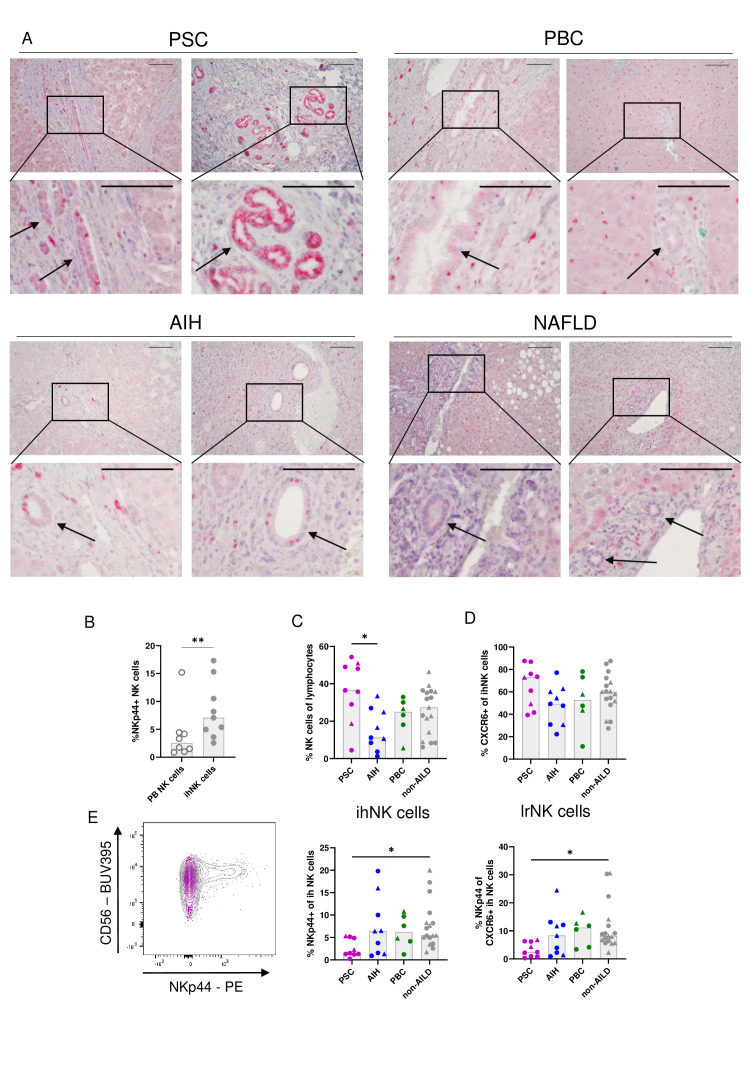
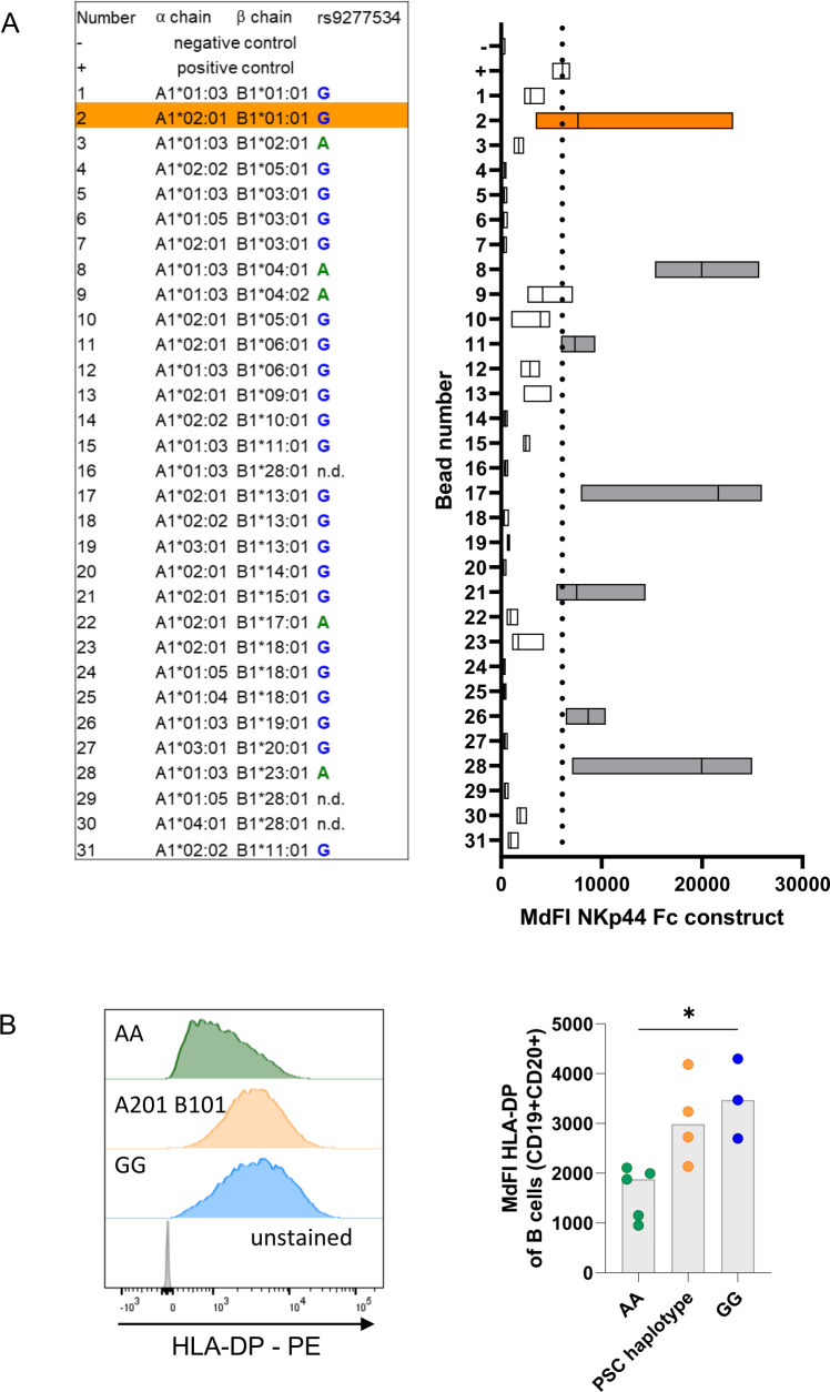
Objective: Primary sclerosing cholangitis (PSC) is characterised by bile duct strictures and progressive liver disease, eventually requiring liver transplantation. Although the pathogenesis of PSC remains incompletely understood, strong associations with HLA-class II haplotypes have been described. As specific HLA-DP molecules can bind the activating NK-cell receptor NKp44, we investigated the role of HLA-DP/NKp44-interactions in PSC.

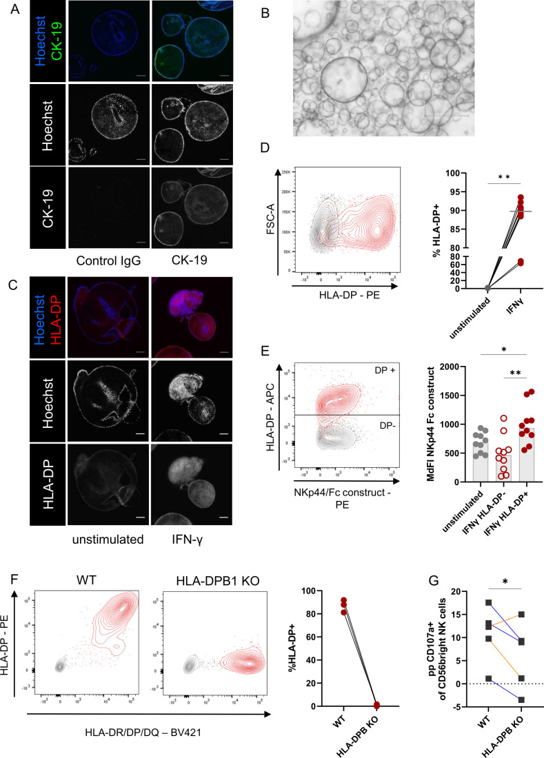
Design: Liver tissue, intrahepatic and peripheral blood lymphocytes of individuals with PSC and control individuals were characterised using flow cytometry, immunohistochemical and immunofluorescence analyses. HLA-DPA1 and HLA-DPB1 imputation and association analyses were performed in 3408 individuals with PSC and 34 213 controls. NK cell activation on NKp44/HLA-DP interactions was assessed in vitro using plate-bound HLA-DP molecules and HLA-DPB wildtype versus knock-out human cholangiocyte organoids.

Results: NKp44+NK cells were enriched in livers, and intrahepatic bile ducts of individuals with PSC showed higher expression of HLA-DP. HLA-DP haplotype analysis revealed a highly elevated PSC risk for HLA-DPA1*02:01~B1*01:01 (OR 1.99, p=6.7×10-50). Primary NKp44+NK cells exhibited significantly higher degranulation in response to plate-bound HLA-DPA1*02:01-DPB1*01:01 compared with control HLA-DP molecules, which were inhibited by anti-NKp44-blocking. Human cholangiocyte organoids expressing HLA-DPA1*02:01-DPB1*01:01 after IFN-γ-exposure demonstrated significantly increased binding to NKp44-Fc constructs compared with unstimulated controls. Importantly, HLA-DPA1*02:01-DPB1*01:01-expressing organoids increased degranulation of NKp44+NK cells compared with HLA-DPB1-KO organoids.

Conclusion: Our studies identify a novel PSC risk haplotype HLA-DP A1*02:01~DPB1*01:01 and provide clinical and functional data implicating NKp44+NK cells that recognise HLA-DPA1*02:01-DPB1*01:01 expressed on cholangiocytes in PSC pathogenesis.

Keywords: AUTOIMMUNE BILIARY DISEASE; IMMUNE-MEDIATED LIVER DAMAGE; IMMUNOGENETICS; IMMUNOLOGY IN HEPATOLOGY; PRIMARY SCLEROSING CHOLANGITIS.


Conflict of interest statement

Competing interests: None declared.

Figures

Figure 1
Figure 1
Upregulation of HLA-DP expression and reduced frequencies of intrahepatic NKp44+NK cells in individuals with PSC. (A) Representative images of immunohistochemical analyses of HLA-DP (permanent red) in PSC, PBC and AIH-affected liver sections and non-AILD controls (NAFLD). Scale bars indicate 100 µm. Arrows point at intrahepatic bile ducts. (NAFLD n=6; PSC n=8, AIH n=6, PBC n=7). (B) Frequencies of NKp44+NK cells in matched peripheral blood (PB) and intrahepatic (ih) NK cells of non-AILD liver tissue. (C) Frequencies of NK cells within intrahepatic (ih) lymphocytes of individuals with PSC, AIH, PBC and non-AILD controls. (D) Frequencies of CXCR6+NK cells within ihNK cells of individuals with PSC, AIH, PBC and non-AILD controls. (E) Expression of NKp44 by intrahepatic (ih; middle panel), liver-resident (lr; right panel) NK cells of individuals with PSC, AIH, PBC and non-AILD controls. Left panel: representative flow cytometry plot (non-AILD—grey, PSC—magenta). Dots represent explant liver samples and resection margins of liver metastases. Triangles represent fresh liver biopsies. Bars indicate the median. Wilcoxon test was used to assess statistical differences between matched PB and liver samples (B). **p=0.0078. Kruskal-Wallis test (Dunn’s post-test) was used to assess statistical differences between PSC and control groups (C–E). (C) *p=0.0190, (E) ih NK cells *p=0.0169; lr NK cells *p=0.0120. AIH, autoimmune hepatitis; AILD, autoimmune liver disease; NAFLD, non-alcoholic fatty liver disease; PBC, primary biliary cholangitis; PSC, primary sclerosing cholangitis.
Figure 2
Figure 2
PSC HLA-DP susceptibility alleles encode for an NKp44-binding HLA-DP molecule (A) Median fluorescence intensity (MdFI) of NKp44 Fc-construct binding (x-axis) to HLA-DP coated beads. Depicted are median (line) and range (boxes) of three individual experiments. The dotted line represents the MdFI of the positive control. Orange: HLA-DPA1*02:01-DPB1*01:01 (consisting of both PSC risk alleles), grey: all other HLA-DP molecules with NKp44/Fc-construct binding above the positive control. Left: Table indicating HLA-DP alleles of the coated beads. Association of the HLA-DP beta-chain alleles with the SNP rs9277534 is marked according to Schöne et al. (B) MdFI of HLA-DP expression on CD3- CD19+CD20+ B cells of healthy donors encoding for HLA-DP haplotypes linked to rs9277534G/G, rs9277534A/A or HLA-DPA1*02:01~DPB1*01:01 rs9277534G/A. Each dot represents results from one donor. Bars indicate the median. Kruskal-Wallis test with Dunn’s post-test was used to assess statistical differences between the groups. *p=0.034. PSC, primary sclerosing cholangitis.
Figure 3
Figure 3
PSC susceptibility haplotype HLA-DP A1*02:01~DPB1*01:01 enables binding to NKp44, leading to activation of NK cells (A) Left panel: representative plots of CD107a expression (y-Axis) of prestimulated NK cells on incubation with plate-bound anti-NKp44 (grey), HLA-DPA1*01:03-DPB1*03:01 (blue), HLA-DPA1*02:01-DPB1*01:01 (orange) or PBS as negative control (black). Right panel: percentages of CD107a+NK cells in the plate-bound degranulation assay after incubation with the respective HLA-DP molecules minus PBS negative control. Wilcoxon-signed rank test was used to assess statistical differences between the different HLA-DP molecules; **p=0.0078. (B) Representative plots (upper panel) and percentages of CD107a+NK cells (lower panel) in a plate-bound degranulation assay after incubation with anti-NKp44 (left) or the respective HLA-DP molecules (middle HLA-DPA1*01:03-DPB1*03:01, right HLA-DPA1*02:01-DPB1*01:01) minus PBS negative controls after preincubation with anti-NKp44 blocking antibody or the isotype control antibodies. Wilcoxon-signed rank test was used to assess statistical differences between the groups. **p=0.0039 (aNKp44); **p=0.0078 (HLA-DPA201-DPB101). (C) MdFI of NKp44 expression on NK cells in the plate-bound degranulation assay on incubation with PBS and plate-bound anti-NKp44 (left), HLA-DPA1*01:03-DPB1*03:01 (middle), and HLA-DPA1*02:01-DPB1*01:01 (right). Lines connect the corresponding conditions of one donor. Wilcoxon-signed rank test was used to assess statistical differences between the groups. **p=0.0039 (aNKp44); p=0.0547 (HLA-DPA201-DPB101).
Figure 4
Figure 4
Increased binding of NKp44/Fc construct to HLA-DP expressing cholangiocyte organoids and reduced degranulation of NK cells towards HLA-DPB1 KO cholangiocyte organoids (A) Immunofluorescence analyses of cholangiocyte organoids for Cytokeratin 19 (CK-19—green) and Hoechst blue (blue). Full staining (right panel) and control IgG (left panel). Scale bar represents 100 µm. (B) Light microscopy images of cholangiocyte organoids showing cystic structures. Scale bar represents 750 µm. (C) Immunofluorescence analyses of cholangiocyte organoids for Hoechst (blue) and HLA-DP PE (red) unstimulated (left panel) and IFN-γ-stimulated (right panel). Scale bar represents 100 µm. (D) Left panel: Representative plot of HLA-DP expression (x-Axis) of unstimulated (grey) and IFNγ-stimulated cholangiocytes from organoids (red). Right panel: Percentage of HLA-DP+cholangiocytes from organoids within unstimulated or IFN-γ-stimulated cultures. n=10, from five donors in three individual experiments. Wilcoxon-signed rank test was used to assess statistical differences between the matched conditions. **p=0.002. (E) Left panel: representative plot of HLA-DP expression (y-Axis) and NKp44/Fc-construct binding (x-axis) of cholangiocytes from organoids unstimulated (grey) or IFN-γ-stimulated (red). Right panel: MdFI of NKp44/Fc-construct binding to cholangiocytes from organoids unstimulated (grey) or IFN-γ-stimulated HLA-DP- populations (red circles) and IFN-γ-stimulated HLA-DP+populations (red dots). Friedman test with Dunn’s post-test was used to assess statistical differences between the groups. *p=0.0417; **p=0.001. (n=10, from four individual donors; performed in three individual experiments). (F) Expression of HLA-DP on WT and HLA-DPB1 KO cholangiocytes from organoids after IFN-γ-stimulation, performed in tree individual experiments. Left: representative plot: HLA-DP (y-axis) and HLA-DR/DQ/DP-expression (x-axis) of WT (left) or HLA-DPB1 KO (right) cholangiocytes from organoids after stimulation with IFN-γ (red) and unstimulated control (grey), right: graph showing frequencies of HLA-DP+cholangiocyte organoid cells. (G) Depicted is the CD107a expression of prestimulated NK cells after co-incubation with IFN-γ-stimulated WT or HLA-DPB1 KO organoids minus PBS negative control. (NK cell donors: autologous NK cells in triplicate—blue lines; HLA/KIR-ligand matched NK cells, one donor in duplicate—orange lines). One-tailed t-test to test the hypothesis that HLA-DPB KO leads to reduced degranulation of NKp44+NK cells, p=0.044.

Similar articles

Cited by

References

    1. Trivedi PJ, Hirschfield GM. Recent advances in clinical practice: epidemiology of autoimmune liver diseases. Gut 2021;70:1989–2003. 10.1136/gutjnl-2020-322362 - DOI - PubMed
    1. Thorpe ME, Scheuer PJ, Sherlock S. Primary Sclerosing cholangitis, the biliary tree, and ulcerative colitis. Gut 1967;8:435–48. 10.1136/gut.8.5.435 - DOI - PMC - PubMed
    1. Karlsen TH, Folseraas T, Thorburn D, et al. . Primary sclerosing cholangitis - a comprehensive review. J Hepatol 2017;67:1298–323. 10.1016/j.jhep.2017.07.022 - DOI - PubMed
    1. Visseren T, Erler NS, Heimbach JK, et al. . Inflammatory conditions play a role in recurrence of PSC after liver transplantation: an international Multicentre study. JHEP Rep 2022;4:100599. 10.1016/j.jhepr.2022.100599 - DOI - PMC - PubMed
    1. Jiang X, Karlsen TH. Genetics of primary Sclerosing cholangitis and pathophysiological implications. Nat Rev Gastroenterol Hepatol 2017;14:279–95. 10.1038/nrgastro.2016.154 - DOI - PubMed

Publication types