

Case Reports
doi: 10.1212/WNL.0000000000207857.
Epub 2023 Oct 3.
Pearls & Oy-sters: Adult-Onset Craniopharyngioma Presenting With Cognitive Dysfunction and Obstructive Hydrocephalus
Affiliations
- PMID: 37788936
- PMCID: PMC10663027
- DOI: 10.1212/WNL.0000000000207857
Item in Clipboard
Case Reports
Pearls & Oy-sters: Adult-Onset Craniopharyngioma Presenting With Cognitive Dysfunction and Obstructive Hydrocephalus
Neurology.
.
Erratum in
-
Corrections to Received Date Information.Neurology. 2024 Jul 9;103(1):e209596. doi: 10.1212/WNL.0000000000209596. Epub 2024 Jun 3. Neurology. 2024. PMID: 38830175 Free PMC article. No abstract available.
No abstract available
Conflict of interest statement
The authors report no relevant disclosures. Go to
Figures
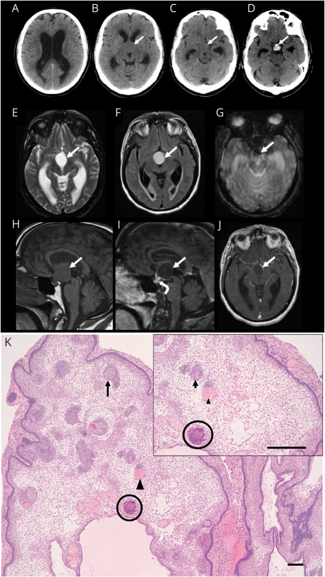
Axial CT images demonstrating hydrocephalus with lateral ventricle dilatation (A) secondary to a suprasellar mass (arrow, B and C) with intrinsic calcification at its base anterior to the dorsum sella (arrow, D). MRI brain axial T2-weighted (E), fluid-attenuated inversion recovery (F), gradient-echo (G), and sagittal T1-weighted (H) images demonstrate a proteinaceous cyst in the suprasellar cistern with rim enhancement on sagittal (I) and axial (J) postcontrast T1-weighted images (solid arrows) and solid enhancement at the base of the lesion (curved arrow, I). Susceptibility artifact on the axial gradient-echo image (arrow, G) corresponds to the calcification at the base of the lesion. Paraffin-embedded, hematoxylin and eosin–stained section (K) reveals a multilobulated mass that is composed of palisading epithelium surrounding a loose stellate reticulum. Whorls of squamous epithelium (arrow), nodules of anucleate squames (arrowhead), and calcifications (circle) are present within the reticulum (scale bar 10 μm).
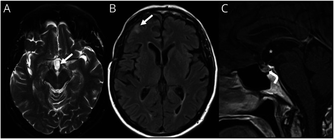
Axial T2-weighted (A) and fluid-attenuated inversion recovery (B) MR brain images and sagittal postcontrast T1-weighted image (C) demonstrating postoperative changes after tumor resection and radiosurgery with resection cavity in suprasellar region (arrow, A; asterisk, C), gliosis in right frontal lobe beneath a craniotomy defect (arrow, B), resolution of hydrocephalus, and normal appearance to the pituitary gland (curved arrow, C).
References
Publication types
MeSH terms
LinkOut - more resources
Full Text Sources
Medical