Case Reports
. 2023 Nov 21;101(21):e2197-e2201.
doi: 10.1212/WNL.0000000000207855. Epub 2023 Oct 3.

Clinical Reasoning: A Woman With Progressive Painless Sequential Monocular Vision Loss

Affiliations
Case Reports

Clinical Reasoning: A Woman With Progressive Painless Sequential Monocular Vision Loss

Joseph Ditrapani et al. Neurology. .

Erratum in

  • Corrections to Received Date Information.
    [No authors listed] [No authors listed] Neurology. 2024 Jul 9;103(1):e209596. doi: 10.1212/WNL.0000000000209596. Epub 2024 Jun 3. Neurology. 2024. PMID: 38830175 Free PMC article. No abstract available.

Abstract

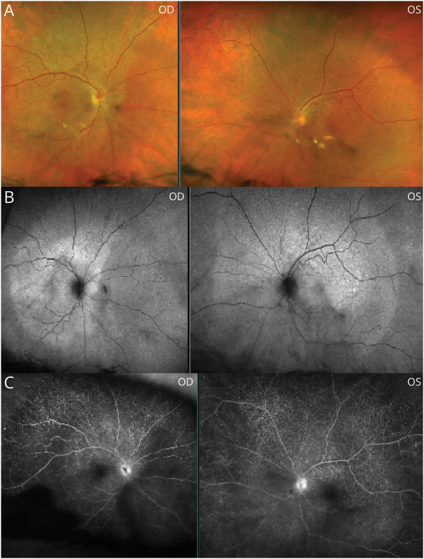
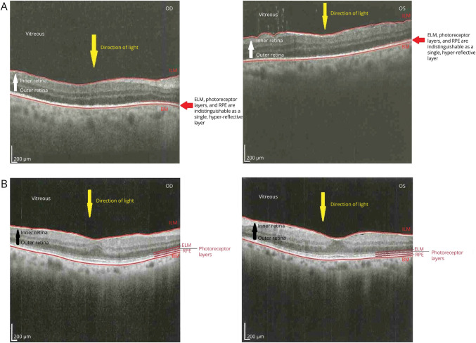
A 68-year-old woman with a history of diabetes mellitus type 2, depression, and migraines presented with painless, acute, consecutive vision loss affecting the right eye for 1 week and the left eye for 2 weeks. Neuro-ophthalmic examination was notable for visual acuities of finger-counting peripherally, a central scotoma, anterior uveitis, vitritis, and placoid macular pigmentary changes in each eye (OU). Proprioception was diminished in the bilateral lower extremities. Optical coherence tomography (OCT) revealed hyper-reflectivity and attenuation of the outer retina OU with normal inner retinal architecture and reflectivity. Fluorescein angiography (FA) demonstrated normal filling of the central retinal arteries with patchy choroidal perfusion in the right eye and targetoid punctate foci of leakage in the macula OU. Before the recognition of intraocular inflammation and findings on OCT and FA, the patient was treated for presumed central retinal artery occlusion at an outside hospital. Additional diagnostic testing at our institution revealed an alternate diagnosis. This case highlights a rare presentation of a well-known disease entity and underscores the importance of avoiding diagnostic anchoring in clinical practice.


Conflict of interest statement

The authors report no relevant disclosures. Go to Neurology.org/N for full disclosures.

Figures

Figure 1
Figure 1. Fundus Photography, Fundus Autofluorescence, and Fluorescein Angiography
Placoid-like macular pigmentary changes are depicted on fundus photography (A) and fundus autofluorescence (B); the sharply delineated margin of the pigmentary change (indicative of the pathologically affected area) is seen. Note also patchy choroidal filling and disc leakage noted on fluorescein angiography (C). OD = right eye; OS = left eye.
Figure 2
Figure 2. OCT Before Treatment and 14 Months Post-treatment
This OCT (A) before treatment with intravenous penicillin demonstrates loss of the delineation of the outer retinal layers—the ELM, photoreceptor layers, and RPE layers—which can be visually differentiated from one another in the healthy eye—and are here supplanted by a single, hyper-reflective layer. These changes correlate with loss of the retina peripheral to the ellipsoid zone on OCT, specifically including the RPE and photoreceptor layers. This OCT (B) from 14 months post-treatment with intravenous penicillin now demonstrates visible delineation between the outer retinal layers, including the ELM, the photoreceptor layers, and the RPE. BM = Bruch membrane; ELM = external limiting membrane; ILM = internal limiting membrane; OCT = optical coherence tomography; RPE = retinal pigment epithelium.

References

    1. Ghanem KG, Ram S, Rice P. The modern epidemic of syphilis. N Engl J Med. 2020;382(9):845-854. doi:10.1056/NEJMra1901593 - DOI - PubMed
    1. Furtado JM, Arantes TE, Nascimento H, et al. . Clinical manifestations and ophthalmic outcomes of ocular syphilis at a time of re-emergence of the systemic infection. Sci Rep. 2018;8(1):12071. doi:10.1038/s41598-018-30559-7 - DOI - PMC - PubMed
    1. Mathew R, Goh B, Westcott M. British Ocular Syphilis Study (BOSS): 2-year national surveillance study of intraocular inflammation secondary to ocular syphilis. Invest Opthalmol Vis Sci. 2014;55(8):5394. doi:10.1167/iovs.14-14559 - DOI - PubMed

Publication types