

Long-term effects of prenatal infection on the human brain: a prospective multimodal neuroimaging study
- PMID: 37789021
- PMCID: PMC10547711
- DOI: 10.1038/s41398-023-02597-x
Long-term effects of prenatal infection on the human brain: a prospective multimodal neuroimaging study
Abstract
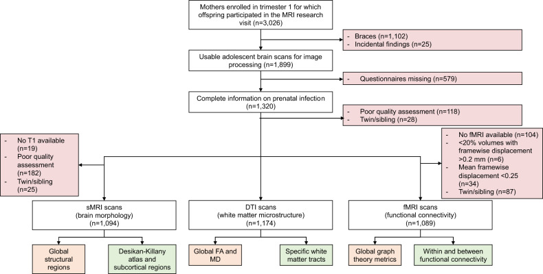
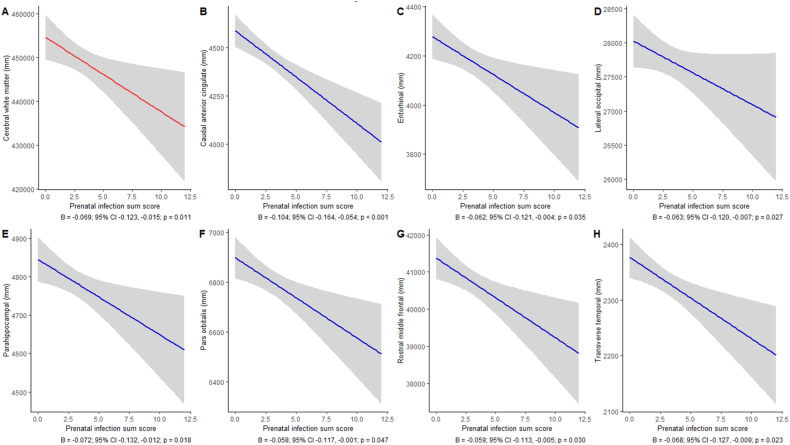
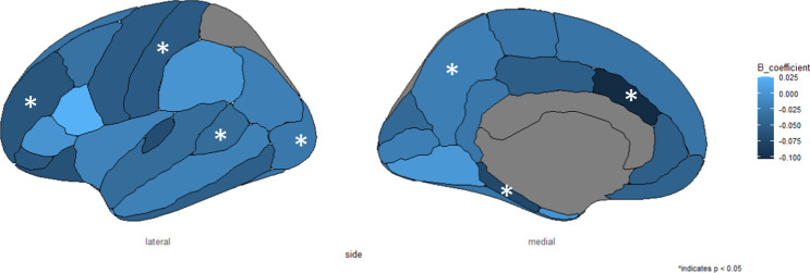
There is convincing evidence from rodent studies suggesting that prenatal infections affect the offspring's brain, but evidence in humans is limited. Here, we assessed the occurrence of common infections during each trimester of pregnancy and examined associations with brain outcomes in adolescent offspring. Our study was embedded in the Generation R Study, a large-scale sociodemographically diverse prospective birth cohort. We included 1094 mother-child dyads and investigated brain morphology (structural MRI), white matter microstructure (DTI), and functional connectivity (functional MRI), as outcomes at the age of 14. We focused on both global and focal regions. To define prenatal infections, we composed a score based on the number and type of infections during each trimester of pregnancy. Models were adjusted for several confounders. We found that prenatal infection was negatively associated with cerebral white matter volume (B = -0.069, 95% CI -0.123 to -0.015, p = 0.011), and we found an association between higher prenatal infection scores and smaller volumes of several frontotemporal regions of the brain. After multiple testing correction, we only observed an association between prenatal infections and the caudal anterior cingulate volume (B = -0.104, 95% CI -0.164 to -0.045, p < 0.001). We did not observe effects of prenatal infection on other measures of adolescent brain morphology, white matter microstructure, or functional connectivity, which is reassuring. Our results show potential regions of interest in the brain for future studies; data on the effect of severe prenatal infections on the offspring's brain in humans are needed.
© 2023. Springer Nature Limited.
Conflict of interest statement
The authors declare no competing interests.
Figures
Similar articles
-
Maternal Immune Activation and Child Brain Development: A Longitudinal Population-Based Multimodal Neuroimaging Study.Biol Psychiatry Cogn Neurosci Neuroimaging. 2025 Feb;10(2):222-235. doi: 10.1016/j.bpsc.2024.10.013. Epub 2024 Nov 2. Biol Psychiatry Cogn Neurosci Neuroimaging. 2025. PMID: 39491788
-
Examining longitudinal associations between prenatal exposure to infections and child brain morphology.Brain Behav Immun. 2024 Jul;119:965-977. doi: 10.1016/j.bbi.2024.05.014. Epub 2024 May 13. Brain Behav Immun. 2024. PMID: 38750701 Free PMC article.
-
Associations of Air Pollution on the Brain in Children: A Brain Imaging Study.Res Rep Health Eff Inst. 2022 Feb;2022(209):1-61. Res Rep Health Eff Inst. 2022. PMID: 36106707 Free PMC article.
-
Neuroimaging effects of prenatal alcohol exposure on the developing human brain: a magnetic resonance imaging review.Acta Neuropsychiatr. 2015 Oct;27(5):251-69. doi: 10.1017/neu.2015.12. Epub 2015 Mar 17. Acta Neuropsychiatr. 2015. PMID: 25780875 Review.
-
Effects of Prenatal Methamphetamine Exposure on the Developing Human Brain: A Systematic Review of Neuroimaging Studies.ACS Chem Neurosci. 2021 Aug 4;12(15):2729-2748. doi: 10.1021/acschemneuro.1c00213. Epub 2021 Jul 23. ACS Chem Neurosci. 2021. PMID: 34297546 Free PMC article.
Cited by
-
The association between maternal immune activation and brain structure and function in human offspring: a systematic review.Mol Psychiatry. 2025 Feb;30(2):722-735. doi: 10.1038/s41380-024-02760-w. Epub 2024 Sep 28. Mol Psychiatry. 2025. PMID: 39342040 Free PMC article.
-
Maternal Immune Activation and Child Brain Development: A Longitudinal Population-Based Multimodal Neuroimaging Study.Biol Psychiatry Cogn Neurosci Neuroimaging. 2025 Feb;10(2):222-235. doi: 10.1016/j.bpsc.2024.10.013. Epub 2024 Nov 2. Biol Psychiatry Cogn Neurosci Neuroimaging. 2025. PMID: 39491788
-
Examining longitudinal associations between prenatal exposure to infections and child brain morphology.Brain Behav Immun. 2024 Jul;119:965-977. doi: 10.1016/j.bbi.2024.05.014. Epub 2024 May 13. Brain Behav Immun. 2024. PMID: 38750701 Free PMC article.
References
-
- Zimmer A, Youngblood A, Adnane A, Miller BJ, Goldsmith DR. Prenatal exposure to viral infection and neuropsychiatric disorders in offspring: a review of the literature and recommendations for the COVID-19 pandemic. Brain Behav Immun. 2021;91:756–70. doi: 10.1016/j.bbi.2020.10.024. - DOI - PMC - PubMed
Publication types
MeSH terms
Grants and funding
LinkOut - more resources
Full Text Sources
