. 2023 Oct 3;13(1):16577.
doi: 10.1038/s41598-023-43563-3.

Effects of tea, catechins and catechin derivatives on Omicron subvariants of SARS-CoV-2

Affiliations

Effects of tea, catechins and catechin derivatives on Omicron subvariants of SARS-CoV-2

Masaharu Shin-Ya et al. Sci Rep. .

Abstract

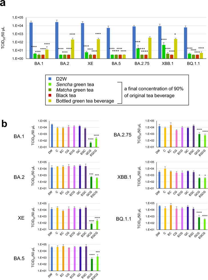
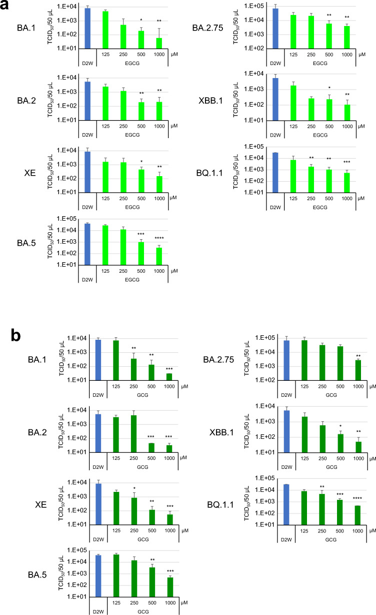
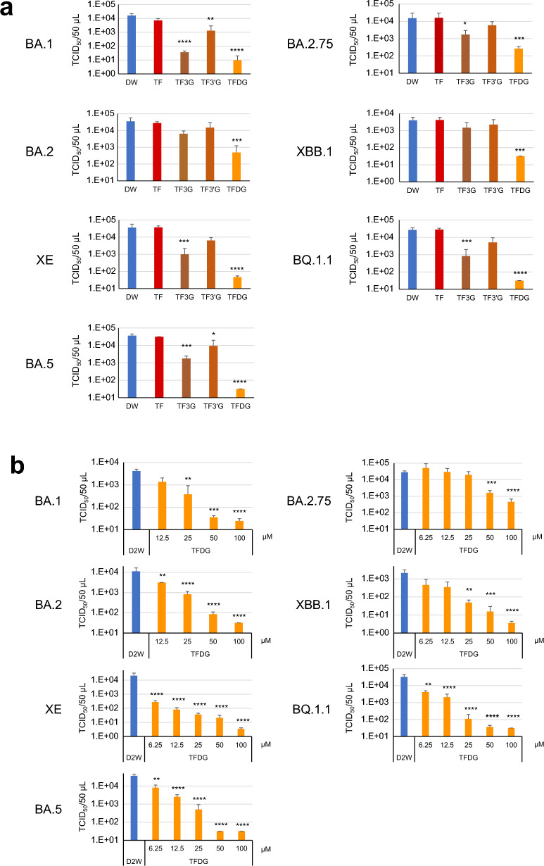
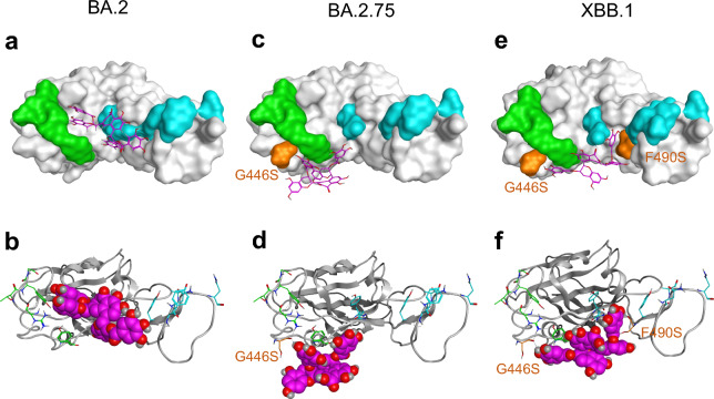
The Omicron subvariants of SARS-CoV-2 have multiple mutations in the S-proteins and show high transmissibility. We previously reported that tea catechin (-)-epigallocatechin gallate (EGCG) and its derivatives including theaflavin-3,3'-di-O-digallate (TFDG) strongly inactivated the conventional SARS-CoV-2 by binding to the receptor binding domain (RBD) of the S-protein. Here we show that Omicron subvariants were effectively inactivated by green tea, Matcha, and black tea. EGCG and TFDG strongly suppressed infectivity of BA.1 and XE subvariants, while effect on BA.2.75 was weaker. Neutralization assay showed that EGCG and TFDG inhibited interaction between BA.1 RBD and ACE2. In silico analyses suggested that N460K, G446S and F490S mutations in RBDs crucially influenced the binding of EGCG/TFDG to the RBDs. Healthy volunteers consumed a candy containing green tea or black tea, and saliva collected from them immediately after the candy consumption significantly decreased BA.1 virus infectivity in vitro. These results indicate specific amino acid substitutions in RBDs that crucially influence the binding of EGCG/TFDG to the RBDs and different susceptibility of each Omicron subvariant to EGCG/TFDG. The study may suggest molecular basis for potential usefulness of these compounds in suppression of mutant viruses that could emerge in the future and cause next pandemic.


Conflict of interest statement

O.M. has received funding support from Japan Science and Technology Agency and ITO EN, ltd, Tokyo, Japan. Other authors declare no COI. The company also provided information of tea, catechins and catechin derivatives, and discussion with authors. The funders had no role in the design of the study, in the interpretation of data, in the writing of the manuscript, or in the decision to publish the results.

Figures

Figure 1
Figure 1
Analysis of inactivation effects of tea and green tea catechins on SARS-CoV-2 Omicron variants. Viruses were treated with the indicated tea (a) or each catechin at the same concentration in bottled green tea beverage (Supplementary Table 1) (b) for 10 s. After dilution, the virus was infected to VeroE6/TMPRSS2 cells to determine TCID50 values as described in the Materials and Methods. Virus titer of each sample (means ± S.D.) is shown (n = 4). ****p < 0.0001, ***p < 0.001, **p < 0.01, and *p < 0.05 vs. Control (distilled water) (Tukey’s multiple comparisons test).
Figure 2
Figure 2
Effect of EGCG and GCG on Omicron subvariants. Omicron subvariants were treated with the indicated concentrations of EGCG (a) or GCG (b) for 10 s. After dilution, virus titers (means ± S.D.) were determined as in Fig. 1 (n = 4). ****p < 0.0001, ***p < 0.001, **p < 0.01, and *p < 0.05 vs. Ctrl (distilled water) (Tukey's multiple comparisons test).
Figure 3
Figure 3
Effect of theaflavins on Omicron subvariants. Viruses were treated with each compound at the same concentration in black tea (a) or with the indicated concentrations of TFDG (b) for 10 s. After dilution, virus titers (means ± S.D.) were determined as in Fig. 1 (n = 4). ****p < 0.0001, ***p < 0.001, **p < 0.01, and *p < 0.05 vs. Ctrl (distilled water) (Tukey’s multiple comparisons test).
Figure 4
Figure 4
EGCG, GCG, and TFDG hampered the interaction between ACE2 and RBD of Omicron BA.1. (a) BA.1 virus and EGCG was mixed, and the mixture was infected to cells (“Mix”), while other cells were pre-treated with EGCG followed by washing and infection by DW-treated BA.1 virus (“Pre”). As a control (DW), DW-treated BA.1 virus was infected to non-treated cells. Virus titers were determined as in Fig. 1. (bd) HRP-RBD was mixed with EGCG, and the mixture was added to ACE2-pre-coated wells (“Mix protocol”) (b,c), and green bars in (d), while other ACE2-pre-coated wells were pretreated with EGCG followed by washing and an addition of DW-treated HRP-RBD (“Pretreatment protocol”) (pink bars in (d)). As a control (DW), DW-treated HRP-RBD was added to ACE2-pre-coated wells. The binding between RBD and ACE2 was evaluated, and % Inhibition for each sample is shown. Values are means ± S.D. (n = 3). ****p < 0.0001 vs. Control (DW), N.S., p > 0.05 vs. Control (DW), +  +  +  +p < 0.0001, between indicated groups, by Tukey’s multiple comparison test.
Figure 5
Figure 5
Structure of EGCG binding with the Omicron RBD of the BA.1 and BA.2.75 lineages. (a) EGCG (Stick model as yellow) on the binding surface of RBD of the BA.1 lineage (Surface model). (b) EGCG (Space-filling model) on the binding surface of RBD of the BA.1 lineage (Skelton model). (c) EGCG (Stick model as yellow) on the binding surface of RBD of the BA.2.75 lineage (Surface model). (d) EGCG (Space-filling model) on the binding surface of RBD of the BA.2.75 lineage (Skelton model). The residues in Patch 1 of RBD are shown as cyan sticks, and those in Patch 2 of RBD are shown as green sticks. The residue (N460K) mutated in the BA.2.75 lineage is shown as orange.
Figure 6
Figure 6
Structural comparison of TFDG binding with the Omicron RBD. (a) TFDG (Stick model as Magenta) on the binding surface of RBD of the BA.2 lineage (Surface model). (b) TFDG (Space-filling model) on the binding surface of RBD of the BA.2 lineage (Skelton model). (c) TFDG (Stick model as Magenta) on the binding surface of RBD of the BA.2.75 lineage (Surface model). (d) TFDG (Space-filling model) on the binding surface of RBD of the BA.2.75 lineage (Skelton model). (e) TFDG (Stick model as Magenta) on the binding surface of RBD of the XBB.1 lineage (Surface model). (f) TFDG (Space-filling model) on the binding surface of RBD of the XBB.1 lineage (Skelton model). The residues in Patch 1 of RBD are shown as cyan sticks, and those in Patch 2 of RBD are shown as green sticks. The G446S and F490S mutated residues are shown as orange.
Figure 7
Figure 7
Inactivation of BA.1 Virus in vitro by Saliva from Volunteers Who Consumed a Candy Containing Tea. Healthy volunteers consumed a placebo candy (without tea) or a candy containing either green tea or black tea for 5 min. Saliva was collected from the volunteers before, or the indicated time after cessation of, the candy consumption. BA.1 virus was treated with each saliva sample for 10 s. After dilution, virus titers were determined by TCID50 method (n = 7 persons). (a) Kinetic change of virus titers for each single person is plotted. (b) Means ± S.D. of virus titers are shown. ****p < 0.0001, *p < 0.05, vs. placebo candy (without tea) group at the same time point. +  +  +  + p < 0.0001, vs. “before” time point of the same groups (Tukey's multiple comparisons test).

References

    1. Viana R, et al. Rapid epidemic expansion of the SARS-CoV-2 Omicron variant in southern Africa. Nature. 2022;603:679–686. doi: 10.1038/s41586-022-04411-y. - DOI - PMC - PubMed
    1. Willett BJ, et al. SARS-CoV-2 Omicron is an immune escape variant with an altered cell entry pathway. Nat. Microbiol. 2022;7:1161–1179. doi: 10.1038/s41564-022-01143-7. - DOI - PMC - PubMed
    1. Hu B, et al. Spike mutations contributing to the altered entry preference of SARS-CoV-2 omicron BA.1 and BA.2. Emerg. Microbes Infect. 2022;11:2275–2287. doi: 10.1080/22221751.2022.2117098. - DOI - PMC - PubMed
    1. Rathnasinghe R, et al. Characterization of SARS-CoV-2 Spike mutations important for infection of mice and escape from human immune sera. Nat. Commun. 2022;13:3921. doi: 10.1038/s41467-022-30763-0. - DOI - PMC - PubMed
    1. Schubert M, et al. Human serum from SARS-CoV-2-vaccinated and COVID-19 patients shows reduced binding to the RBD of SARS-CoV-2 Omicron variant. BMC Med. 2022;20:102. doi: 10.1186/s12916-022-02312-5. - DOI - PMC - PubMed

Publication types