. 2023 Oct 3;56(1):52.
doi: 10.1186/s40659-023-00458-x.

Focal ischemic stroke modifies microglia-derived exosomal miRNAs: potential role of mir-212-5p in neuronal protection and functional recovery

Affiliations

Focal ischemic stroke modifies microglia-derived exosomal miRNAs: potential role of mir-212-5p in neuronal protection and functional recovery

Si-Si Li et al. Biol Res. .

Abstract

Background: Ischemic stroke is a severe type of stroke with high disability and mortality rates. In recent years, microglial exosome-derived miRNAs have been shown to be promising candidates for the treatment of ischemic brain injury and exert neuroprotective effects. Mechanisms underlying miRNA dysregulation in ischemic stroke are still being explored. Here, we aimed to verify whether miRNAs derived from exosomes exert effects on functional recovery.

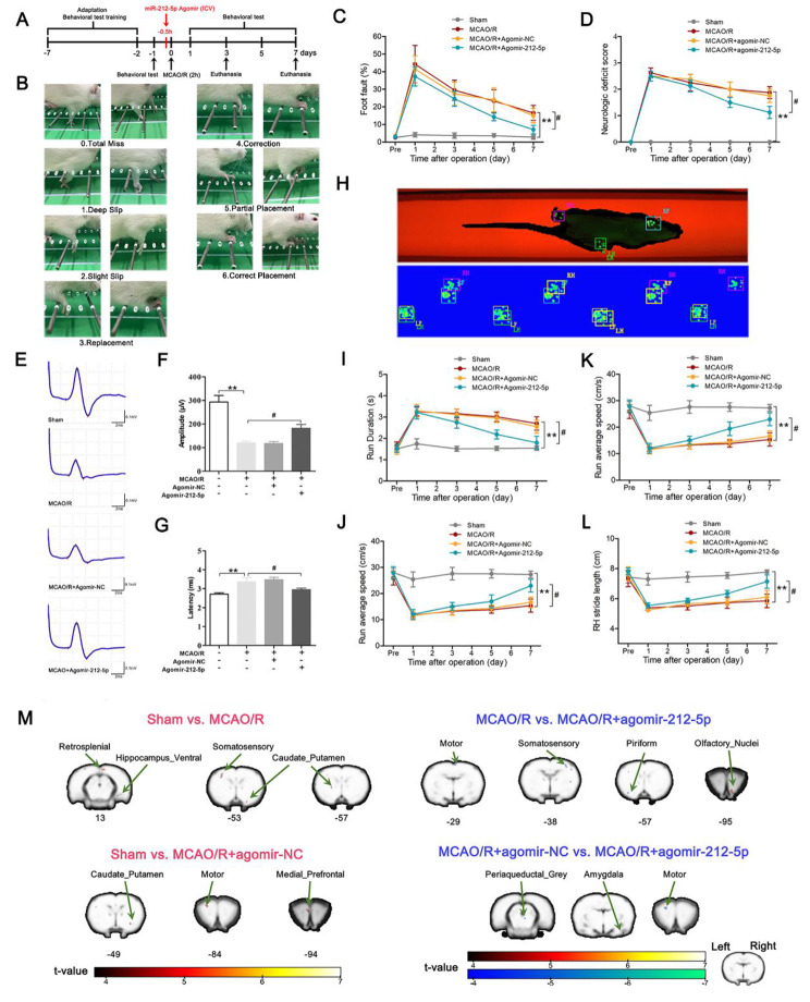
Methods: MiR-212-5p agomir was employed to upregulate miR-212-5p expression in a rat model of middle cerebral artery occlusion/reperfusion (MCAO/R) as well as an oxygen-glucose deprivation/reoxygenation (OGD/R) in vitro. Western blot analysis, qRT-PCR and immunofluorescence staining and other methods were applied to explore the underlying mechanisms of action of miR-212-5p.

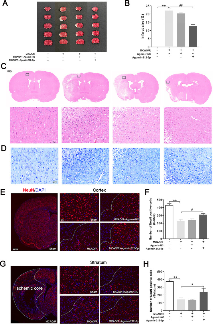
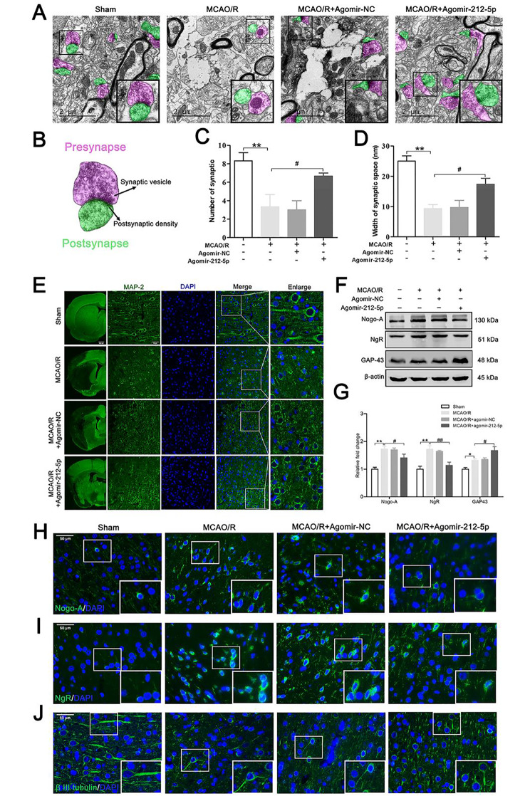
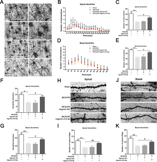
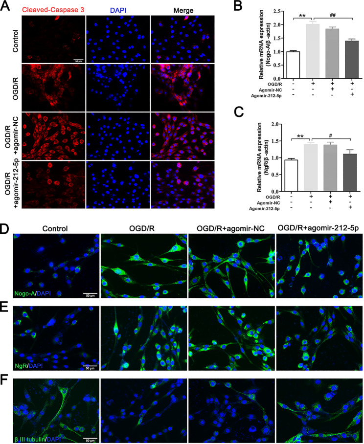
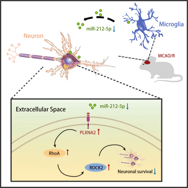
Results: The results of our study found that intervention with miR-212-5p agomir effectively decreased infarct volume and restored motor function in MCAO/R rats. Mechanistically, miR-212-5p agomir significantly reduced the expression of PlexinA2 (PLXNA2). Additionally, the results obtained in vitro were similar to those achieved in vivo.

Conclusion: In conclusion, the present study indicated that PLXNA2 may be a target gene of miR-212-5p, and miR-212-5p has great potential as a target for the treatment and diagnosis of ischemic stroke.

Keywords: Exosomal; Ischemic stroke; MiR-212-5p; Microglial; Neuronal protection; PlexinA2.


Conflict of interest statement

The authors declare that they have no competing interests.

Figures

Fig. 1
Fig. 1
Measurement of pro- and anti‐inflammatory cytokine levels at 6 h, 1, 3, 5 and 7 days postsurgery. A-E Proinflammatory cytokines released by microglia (IL‐1β, IL-6, INOS, CD32 and CD86). F-H Anti-inflammatory cytokines released by microglia (anti‐inflammatory cytokines TGF-β, IL‐10 and CD206). I Double immunofluorescence staining was applied to examine the expression of CD86 (in green) and Iba1 (in red) in the ischemic penumbra of the cortex at different time points. J Double immunofluorescence staining was performed to examine the expression of CD206 (in green) and Iba1 (in red) at different time points. Scale bar = 50 μm. The data are presented as the means ± SEM (n = 5 per group). *P < 0.05 and **P < 0.01 compared with the sham group
Fig. 2
Fig. 2
MiRNA sequencing of microglial exosomes 3 days after MCAO/R and qRT–PCR validation. A, B Characterisation of microglial exosomes using nanoparticle tracking analysis and transmission electron microscopy scanning. Scale bar = 200 nm. C The exosome markers CD9, CD63, and CD81 were detected using western blot analysis. D Microglia-derived exosomes were detected by western blot analysis using the microglia marker CD11b. E Heatmap showing the levels of miRNAs in microglial exosomes at 3 days after MCAO/R. F Expression of miR-30c-5p, miR-126a-5p, miR-128-3p, miR-212-5p and miR-1949 in the ischemic penumbra of the cortex at 3 days after MCAO/R was determined using qRT–PCR. G Expression levels of the miRNA-targeting genes PLXNA2, PTEN and FOXO3 in the ischemic penumbra of the cortex at 3 days after MCAO/R. H The target sites of miR-212-5p in PLXNA2 mRNA 3’ untranslated region (3’UTR). I Map of the pmirGLO luciferase reporter vector. J Dual luciferase assays revealed the binding of miR-212-5p to the 3’UTR of PLXNA2. The data are presented as the means ± SEM (n = 5 per group). *P < 0.05 and **P < 0.01 compared with the sham group. #P < 0.05 compared with the miR-NC group
Fig. 3
Fig. 3
Treatment with agomir-212-5p decreased neurological deficits and improved locomotor activity 7 days after MCAO/R. A The flow chart of the experiment. B Schematic of the scoring criteria for the foot fault test. C Foot fault test results. D Neurological deficit scores were evaluated using the Zea Longa 5-point scheme. E Illustrative waveforms of motor evoked potential (MEP) for each group. F The amplitude of MEP for each group. G The latency of MEP for each group. n = 6 per group. H Schematic representation of the CatWalk gait analysis. I Running duration (s). J Average running speed (cm/s). K Right forelimb stride length (cm). L Right hindlimb stride length (cm). The data are presented as the means ± SEM (n = 8 per group). M Results of functional connectivity analysis among four groups. *P < 0.05 and **P < 0.01 compared with the sham group, #P < 0.05 and ##P < 0.01 compared with the MCAO/R group
Fig. 4
Fig. 4
Agomir-212-5p reduces the infarct volume and mitigates neuronal apoptosis 7 days after MCAO/R. A Brain sections were stained with TTC to visualise the ischemic lesions at 7 days after MCAO/R. The white area shows the infarct core. B Quantitative analysis of the percentage of the infarct area. C H&E staining. Scale bars = 1000 μm (low magnification) and 50 μm (high magnification). D Nissl staining. Scale bars = 50 μm. E, F Immunofluorescence staining for NeuN showing the survival of neurons in the peri-infarct cortex 7 days after MCAO/R. G, H The numbers of NeuN + viable neurons in the peri-infarct striatum. The data are presented as the means ± SEM (n = 3 per group). *P < 0.05 and **P < 0.01 compared with the sham group, #P < 0.05 and ##P < 0.01 compared with the MCAO/R group
Fig. 5
Fig. 5
Agomir-212-5p inhibited the PLXNA2 and RhoA/ROCK2 in the ischemic penumbra at 3 days following MCAO/R. A The asterisks denote the observation site. B PLXNA2, RhoA and ROCK2 mRNA expression in the ipsilateral brain was analysed using qRT–PCR. C Representative images of western blots for PLXNA2, RhoA and ROCK2 in each group. β-Actin was detected as an internal reference. D Quantitative analysis of the western blot results. E Immunofluorescence staining for the PLXNA2, RhoA and ROCK2 proteins in neurons from each group. Scale bar = 50 μm. The data are presented as the means ± SEM (n = 4–6 per group). *P < 0.05 and **P < 0.01 compared with the sham group, #P < 0.05 and ##P < 0.01 compared with the MCAO/R group
Fig. 6
Fig. 6
MiR-212-5p promotes synaptic plasticity and attenuates axon degeneration at 7 days following MCAO/R. A Ultrastructures of the synapses in the ischemic penumbra analysed using TEM. Scale bar = 2 μm. B Schematic of the presynaptic (violet) and postsynaptic (green) structures. C The number of synapses in the ischemic penumbra of the cortex in each group. D Width of the synaptic space (nm). n = 3 per group. E Immunofluorescence staining shows MAP-2 (in green) expression in the ischemic penumbra of the cortex. Nuclei were stained with DAPI and are visualised in blue. Scale bar = 50 μm. F Representative images of western blots for Nogo-A, NgR, and GAP-43. G Quantitative analysis of the western blot results. n = 4–6 per group. H-J Immunofluorescence staining was performed to show the presence of Nogo-A, NgR and β III tubulin in the ischemic penumbra of the cortex. Nuclei were stained with DAPI and are visualised in blue. Scale bar =, 50 μm. The data are presented as the means ± SEM. *P < 0.05 and **P < 0.01 compared with the sham group, #P < 0.05 and ##P < 0.01 compared with the MCAO/R group
Fig. 7
Fig. 7
MiR-212-5p promoted dendritic spine formation and dendritic growth at 7 days following MCAO/R. A Representative Golgi-Cox-stained photomicrograph of brains from rats subjected to MCAO/R. Scale bar = 50 μm and 20 μm. B The apical dendritic intersections were counted at increasing distances from the centre of the soma. C The total number of intersections in the apical dendrites. D The basal dendritic intersections were counted at increasing distances from the centre of the soma. E The total number of intersections for the basal dendrites. F The total number of branches of apical dendrites. G The total number of branches of basal dendrites. H Graphs of dendritic spines on apical dendrites. Scale bar = 10 μm. I Density of dendritic spines along apical dendrites. J Graphs of dendritic spines on basal dendrites. Scale bar = 10 μm. K Density of dendritic spines along basal dendrites. The data are presented as the means ± SEM (n = 3 per group). *P < 0.05 and **P < 0.01 compared with the sham group, #P < 0.05 and ##P < 0.01 compared with the MCAO/R group
Fig. 8
Fig. 8
Agomir-212-5p suppressed the PLXNA2 and RhoA/ROCK2 in PC12 cells following OGD/R. A Representative photographs of western blots showing PLXNA2, RhoA and ROCK2 levels in each group. β-Actin was used as an internal reference. B Quantitative analysis of the western blot results. C-E Immunofluorescence staining results for the PLXNA2, RhoA and ROCK2 (in green) proteins in neurons in each group. Nuclei were stained with DAPI and are visualised in blue. Scale bar = 50 μm. F-H qRT–PCR was performed to assess the mRNA levels of PLXNA2, RhoA and ROCK2 in each group. The data are presented as the means ± SEM (n = 4–6 per group). *P < 0.05 and **P < 0.01 compared with the sham group, #P < 0.05 and ##P < 0.01 compared with the MCAO/R group
Fig. 9
Fig. 9
MiR-212-5p reduces neuronal apoptosis and attenuates axonal degeneration in PC12 cells following OGD/R. A Cleaved-Caspase 3 immunofluorescence staining (in green). Nuclei were stained with DAPI and are visualised in blue. Scale bar = 50 μm. B, C Nogo-A and NgR gene expression levels in PC12 cells. Gene expression levels were examined using qRT–PCR and normalised to β-actin. D-F Immunofluorescence staining was conducted to reveal the presence of Nogo-A, NgR and β III tubulin in PC12 cells. Nuclei were stained with DAPI and are visualised in blue. Scale bar = 50 μm. The data are presented as the means ± SEM (n = 5 per group). *P < 0.05 and **P < 0.01 compared with the sham group, #P < 0.05 and ##P < 0.01 compared with the MCAO/R group
Fig. 10
Fig. 10
A schematic diagram showing the potential molecular mechanism by which the miR-212-5p agomir exerts a neuroprotective effect to alleviate ischemic neuronal damage

References

    1. Hankey GJ, Stroke Lancet. 2017;389(10069):641–54. doi: 10.1016/S0140-6736(16)30962-X. - DOI - PubMed
    1. Della-Morte D, Guadagni F, Palmirotta R, Testa G, Caso V, Paciaroni M, Abete P, Rengo F, Ferroni P, Sacco RL, Rundek T. Genetics of ischemic stroke, stroke-related risk factors, stroke precursors and treatments. Pharmacogenomics. 2012;13(5):595–613. doi: 10.2217/pgs.12.14. - DOI - PubMed
    1. Donnan GA, Fisher M, Macleod M, Davis SM. Stroke Lancet. 2008;371(9624):1612–23. doi: 10.1016/S0140-6736(08)60694-7. - DOI - PubMed
    1. Dirnagl U. Pathobiology of injury after stroke: the neurovascular unit and beyond. Ann N Y Acad Sci. 2012;1268:21–5. doi: 10.1111/j.1749-6632.2012.06691.x. - DOI - PubMed
    1. Radak D, Katsiki N, Resanovic I, Jovanovic A, Sudar-Milovanovic E, Zafirovic S, Mousad SA, Isenovic ER. Apoptosis and Acute Brain Ischemia in ischemic stroke. Curr Vasc Pharmacol. 2017;15(2):115–22. doi: 10.2174/1570161115666161104095522. - DOI - PubMed