

Post Traumatic Frontal Sinus Mucocele with Subcutaneous Extension: A Case Report and Literature Review
- PMID: 37789831
- PMCID: PMC10543422
- DOI: 10.2147/IMCRJ.S436224
Post Traumatic Frontal Sinus Mucocele with Subcutaneous Extension: A Case Report and Literature Review
Abstract
Background: Langenbach (1820) first described paranasal sinus mucoceles under the name of hydatids. Roulette (1909) introduced the name mucocele. Paranasal sinus mucocele is the accumulation of mucus secretions and exfoliated epithelium in the sinuses, causing enlargement of the sinus walls. It is considered a cystic, dilatation-eroding lesion. However, the mucocele often occurs as a localized mass, causing bone erosion and displacement of surrounding structures. If left untreated, a nearby mucocele in the brain can become infected and lead to death. Frontal sinuses are often involved; sphenoid, ethmoid, and maxillary mucoceles are rare. Mucoceles usually result from sinus ostium obstruction due to infection, fibrosis, inflammation, trauma, surgery, or obstruction by tumors such as osteomas. Of all causes, patients most often present with cranio-facial trauma (82.97%) and the most common mechanism is human aggression (90.85%).
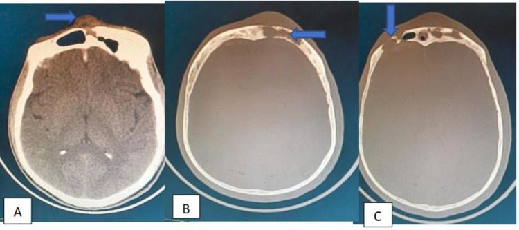
Case presentation: This 30-year-old male patient presented with a frontal head swelling of one year duration that started after he sustained a stick injury on the frontal head one year ago, and he has an associated frontal headache for one year. There was a 4x5cm frontal, firm, palpable, non-tender lesion extending from the nasion to the frontal head. On the brain CT scan, there was frontal bone erosion at multiple sites with partial frontal sinus opacity, an externally growing mass, and an old frontal sinus fracture noted. Bifrontal craniotomy and bilateral frontal sinus cranialization were done, and the patient was discharged on the third day and seen a month later with complete improvement from headache and swelling.
Conclusion: The incidence and pathophysiology of posttraumatic frontal sinus mucoceles are not known yet. The surgical management of mucocele demand a multidisciplinary team involving neurosurgeons, ear nose and throat surgeons, oral and maxillofacial surgeons, ophthalmologists and plastic and reconstructive surgeons. By treating the primary cause, frontal sinus fracture at contact, this case report aims to raise awareness of and prevent frontal sinus mucocele and related complications.
Keywords: cranialization; frontal sinus; mucocele; post-traumatic; subcutaneous extension.
© 2023 Ali.
Conflict of interest statement
The author reports no conflict of interest in this work.
Figures


References
Publication types
LinkOut - more resources
Full Text Sources
