. 2023 Sep 15:14:1096969.
doi: 10.3389/fneur.2023.1096969. eCollection 2023.

Novel variants in the CLCN4 gene associated with syndromic X-linked intellectual disability

Affiliations

Novel variants in the CLCN4 gene associated with syndromic X-linked intellectual disability

Sinan Li et al. Front Neurol. .

Abstract

Objective: The dysfunction of the CLCN4 gene can lead to X-linked intellectual disability and Raynaud-Claes syndrome (MRXSRC), characterized by severe cognitive impairment and mental disorders. This study aimed to investigate the genetic defects and clinical features of Chinese children with CLCN4 variants and explore the effect of mutant ClC-4 on the protein expression level and subcellular localization through in vitro experiments.

Methods: A total of 401 children with intellectual disabilities were screened for genetic variability using whole-exome sequencing (WES). Clinical data, including age, sex, perinatal conditions, and environmental exposure, were collected. Cognitive, verbal, motor, and social behavioral abilities were evaluated. Candidate variants were verified using Sanger sequencing, and their pathogenicity and conservation were analyzed using in silico prediction tools. Protein expression and localization of mutant ClC-4 were measured using Western blotting (WB) and immunofluorescence microscopy. The impact of a splice site variant was assessed with a minigene assay.

Results: Exome analysis identified five rare CLCN4 variants in six unrelated patients with intellectual disabilities, including two recurrent heterozygous de novo missense variants (p.D89N and p.A555V) in three female patients, and two hemizygous missense variants (p.N141S and p.R694Q) and a splicing variant (c.1390-12T > G) that are maternally inherited in three male patients. The p.N141S variant and the splicing variant c.1390-12(T > G were novel, while p.R694Q was identified in two asymptomatic heterozygous female patients. The six children with CLCN4 variants exhibited a neurodevelopmental spectrum disease characterized by intellectual disability (ID), delayed speech, autism spectrum disorders (ASD), microcephaly, hypertonia, and abnormal imaging findings. The minigene splicing result indicated that the c.1390-12T > G did not affect the splicing of CLCN4 mRNA. In vitro experiments showed that the mutant protein level and localization of mutant protein are similar to the wild type.

Conclusion: The study identified six probands with CLCN4 gene variants associated with X-linked ID. It expanded the gene and phenotype spectrum of CLCN4 variants. The bioinformatic analysis supported the pathogenicity of CLCN4 variants. However, these CLCN4 gene variants did not affect the ClC-4 expression levels and protein location, consistent with previous studies. Further investigations are necessary to investigate the pathogenetic mechanism.

Keywords: CLCN4 gene; MRXSRC; intellectual disability; missense variant; splice site variant.


Conflict of interest statement

The authors declare that the research was conducted in the absence of any commercial or financial relationships that could be construed as a potential conflict of interest.

Figures

Figure 1
Figure 1
Clinical findings of affected individuals. (A) Pedigrees of influenced families and variants of the probands. Pedigrees of six unrelated families with CLCN4 variants. The probands are indicated by arrows. I and II indicate the first and second generations, respectively. Squares represent male individuals, circles represent female individuals, filled symbols indicate affected individuals, open symbols indicate healthy individuals, circles with dots indicate asymptomatic carriers, and triangles represent spontaneously aborted fetuses. Number 4 represents the number of abortions. (B) MRI of four patients. P1: Delayed myelination. P3: A widened bilateral frontal-temporal extra brain space and an enlarged left ventricle (indicated by red arrows). P4: Brain dysplasia: small cranial volume; slightly thin posterior of the corpus callosum; widened ventricles and bilateral lateral fissure pools. P6: Possible right temporal arachnoid cyst and slightly wide left temporal subcranial plate gap.
Figure 2
Figure 2
Genetic bioinformatics analysis. (A) Gene sequencing of the six patients with CLCN4 variants. The probands show a nucleotide change (red arrow). Two peaks show a heterozygous nucleotide change and a single peak shows a hemizygous nucleotide change (A, adenosine; G, guanine; C, cytosine; T, thymidine). (B) The variants identified in this study are shown in red. B to Q represent transmembrane and intramembrane domains (green domains represent helical transmembranes and yellow domains represent helical-intramembranes). Multiple-sequence alignment confirms the evolutionary conservation of missense variants in the animal kingdom (CD, cytoplasmic domain; CBS, cystathionine-beta-synthase domains).
Figure 3
Figure 3
Western blot analysis of wild-type and mutated ClC-4. (A) The protein levels of ClC-4 WT, D89N, and R694Q variants in HEK293 cell. (B) Quantification analysis of the ClC-4 WT, D89N, and R694Q variants. (C) The protein levels of ClC-4 WT, A555V, and N141S variants in HEK293 cell. (D) Quantification analysis of the ClC-4 WT, A555V, and N141S variants. Western blot analysis of the HEK293T cell expressing wild-type and mutant ClC-4. The proteins are expressed at similar levels. Differences between groups were calculated using an unpaired t-test with GraphPad Prism software. All experiments were repeated at least three times, and data were presented as the mean ± SD. It was not statistically significant (P > 0.05). All lanes are taken from the same gel and blot.
Figure 4
Figure 4
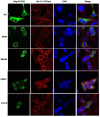
Immunofluorescence analysis of wild-type and mutated ClC-4. Subcellular localization of human ClC-4 variants expressed in HEK293T cells. ClC-4 wildtype and the four mutant proteins localize mainly to the ER. The mutant ClC-4 did not seem to alter the subcellular localization of ClC-4.
Figure 5
Figure 5
Minigene analysis of the novel splicing variant c.1390-12T > G. RT-PCR amplification for aberrant splicing transcripts, agarose gel separation, and subsequent Sanger sequencing was conducted. They were confirmed by bidirectional sequencing. RT-PCR products were analyzed by agarose gel electrophoresis, and no size variants were observed. Sanger sequencing demonstrates no skipping exon.

References

    1. He H, Guzman RE, Cao D, Sierra-Marquez J, Yin F, Fahlke C, et al. . The molecular and phenotypic spectrum of CLCN4-related epilepsy. Epilepsia. (2021) 62:1401–15. 10.1111/epi.16906 - DOI - PubMed
    1. Xu X, Lu F, Zhang L, Li H, Du S, Tang J. Novel CLCN4 variant associated with syndromic X-linked intellectual disability in a Chinese girl: a case report. BMC Pediatr. (2021) 21:384. 10.1186/s12887-021-02860-4 - DOI - PMC - PubMed
    1. Zhou P, He N, Zhang J-W, Lin Z-J, Wang J, Yan L-M, et al. . Novel mutations and phenotypes of epilepsy-associated genes in epileptic encephalopathies. Genes Brain Behav. (2018) 17:e12456. 10.1111/gbb.12456 - DOI - PubMed
    1. Palmer EE, Stuhlmann T, Weinert S, Haan E, van Esch H, Holvoet M, et al. . De novo and inherited mutations in the X-linked gene CLCN4 are associated with syndromic intellectual disability and behavior and seizure disorders in males and females. Mol Psychiatry. (2018) 23:222–30. 10.1038/mp.2016.135 - DOI - PMC - PubMed
    1. Hu H, Haas SA, Chelly J, van Esch H, Raynaud M, de Brouwer AP, et al. . X-exome sequencing of 405 unresolved families identifies seven novel intellectual disability genes. Mol Psychiatry. (2016) 21:133–48. 10.1038/mp.2014.193 - DOI - PMC - PubMed

LinkOut - more resources