[Preprint]. 2023 Sep 21:2023.09.21.558875.
doi: 10.1101/2023.09.21.558875.

The Inflammasome Pathway is Activated by Dengue Virus Non-structural Protein 1 and is Protective During Dengue Virus Infection

Affiliations

The Inflammasome Pathway is Activated by Dengue Virus Non-structural Protein 1 and is Protective During Dengue Virus Infection

Marcus P Wong et al. bioRxiv. .

Update in

Abstract

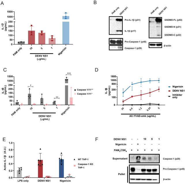
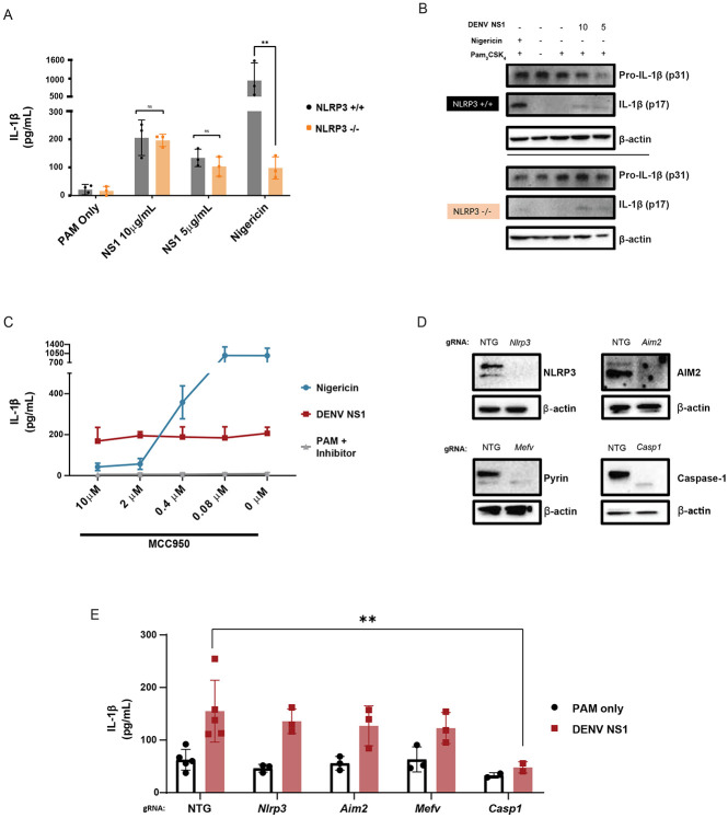
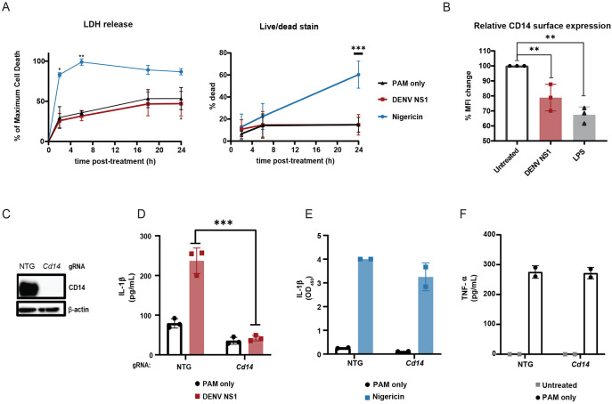
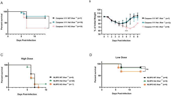
Dengue virus (DENV) is a medically important flavivirus causing an estimated 50-100 million dengue cases annually, some of whom progress to severe disease. DENV non-structural protein 1 (NS1) is secreted from infected cells and has been implicated as a major driver of dengue pathogenesis by inducing endothelial barrier dysfunction. However, less is known about how DENV NS1 interacts with immune cells and what role these interactions play. Here we report that DENV NS1 can trigger activation of inflammasomes, a family of cytosolic innate immune sensors that respond to infectious and noxious stimuli, in mouse and human macrophages. DENV NS1 induces the release of IL-1β in a caspase-1 dependent manner. Additionally, we find that DENV NS1-induced inflammasome activation is independent of the NLRP3, Pyrin, and AIM2 inflammasome pathways, but requires CD14. Intriguingly, DENV NS1-induced inflammasome activation does not induce pyroptosis and rapid cell death; instead, macrophages maintain cellular viability while releasing IL-1β. Lastly, we show that caspase-1/11-deficient, but not NLRP3-deficient, mice are more susceptible to lethal DENV infection. Together, these results indicate that the inflammasome pathway acts as a sensor of DENV NS1 and plays a protective role during infection.


Conflict of interest statement

Declaration of Interests R.E.V. consults for Ventus Therapeutics and X-biotix Therapeutics.

Figures

Figure 1.
Figure 1.. DENV NS1 can activate the inflammasome.
(A) BMDMs were primed with PAM3CSK4 (1μg/mL) for 17 h and then treated with DENV2 NS1 at indicated concentrations, treated with 5μM nigericin, or left untreated (PAM only). IL-1β levels in the supernatant after 2h (nigericin) or 24h (NS1 and PAM only) were measured by ELISA. *p<0.05 as determined by one-way ANOVA with Dunn’s multiple comparison correction. (B) Representative Western blots of BMDM cell lysates after priming with PAM3CSK4 (1μg/mL) for 17h and treatment with 10ug/mL DENV2 NS1 (DENV NS1) or PAM3CSK4 treatment for 24h without NS1 treatment (PAM only). (C) WT and Casp1/11−/− BMDMs were primed with PAM3CSK4 (1μg/mL) for 17h and then treated with DENV2 NS1 at the indicated concentrations, nigericin (5μM), or medium (PAM only). IL-1β levels in the supernatant after 2h (Nigericin) or 24h (NS1 and PAM only) were measured by ELISA. *p<0.05, **p<0.01. Statistical significance was determined using two-way ANOVA followed by multiple t-tests using Holm-Sidak correction. (D) BMDMs were primed with PAM3CSK4 (1μg/mL) for 17h and then pre-treated with Ac-YVAD-cmk at the indicated concentrations before addition of DENV2 NS1 (10μg/mL), nigericin (5μM), or medium (Inhibitor only). IL-1β levels in supernatant 2h (Nigericin) or 24h (NS1 and PAM only) were measured by ELISA. (E) WT or Casp-1−/− THP-1 human monocytes were differentiated into macrophages in 10ng/mL PMA and primed with medium or LPS for 4h. Primed macrophages were treated with DENV NS1 (10μg/mL) or left untreated (LPS only). Eighteen hours later, supernatants were collected. Cells were stimulated with 5μM nigericin for 2h as a positive control. Bioactive IL-1β in supernatants was measured using HEK-Blue IL-1R reporter cells. (F) Representative Western blots of THP-1 macrophage cell lysates after priming with PAM3CSK4 (100ng/mL) for 17h and treatment with DENV2 NS1 at indicated concentrations (μg/mL), treatment with 5μM nigericin, or no treatment for 24h. The data are shown as the mean ± standard deviation (SD) of 3 independent experiments (A, C-D) or 4-6 independent experiments (E) or a representative image taken from 2 biological replicates (B,F).
Figure 2.
Figure 2.. DENV NS1-induced inflammasome activation is NLPR3-independent.
(A) WT and Nlrp3 −/− BMDMs were primed with PAM3CSK4 (1μg/mL) for 17h and then treated with DENV2 NS1 at indicated concentrations, nigericin (5μM), or medium (PAM only). IL-1β levels in supernatant 2h (nigericin) or 24h (NS1 and PAM only) were measured by ELISA. *p<0.05, **p<0.01. Statistical significance was determined using two-way ANOVA followed by multiple t-tests with Holm-Sidak correction. (B) Representative Western blots of cell lysates from WT and Nlrp3−/− BMDMs after priming with PAM3CSK4 (1μg/mL) for 17h and treatment with DENV2 NS1 (10 or 5 μg/mL), treatment with nigericin (5μM), or no treatment for 24h. (C) BMDMs were primed with PAM3CSK4 (1μg/mL) for 17h and then pre-treated with MCC950 at the indicated concentrations before addition of DENV2 NS1 (10μg/mL), nigericin (5μM), or medium (Inhibitor only). IL-1β levels in the supernatant after 2h (Nigericin) or 24h (NS1 and PAM only) were measured by ELISA. (D) Representative Western blots of cell lysates from BMDMs nucleofected with Cas9-gRNA ribonuclear protein complexes to knock out the indicated genes. Two gRNAs per gene were used per nucleofection. NTG = non-targeting guide. (E) Knockout BMDMs from (D) were primed with PAM3CSK4 (1μg/mL) for 17h and treated with DENV2 NS1 (10μg/mL) or left untreated for 48h. *p<0.05. Statistical significance was determined using two-way ANOVA followed by multiple t-tests with Holm-Sidak correction. The data are shown as the mean ± SD of 3 biological replicates (A,C), a representative image taken from 2 biological replicates (B,D), or data pooled from 5 independent experiments with 3 biological replicates per guide (E).
Figure 3.
Figure 3.. DENV NS1 induces inflammasome activation in macrophages in a CD14-dependent manner without inducing cell death.
(A-B) BMDMs were primed with PAM3CSK4 (1μg/mL) for 17h and then treated with DENV2 NS1 (10μg/mL), nigericin (5μM), or medium (PAM only). (A) At the indicated timepoints, supernatants were assessed for lactase dehydrogenase (LDH) levels as a proxy for cell death. LDH levels were calculated as a percentage of maximum LDH release. (B) Cells were stained using a LIVE/DEAD Fixable Far Red stain and analyzed by flow cytometry. *p<0.05 **p<0.01 ***p<0.001. Statistical significance was determined using two-way ANOVA with Dunnetts’s multiple comparison test. (C) BMDMs were primed with PAM3CSK4 (1μg/mL) for 17h and then treated with DENV2 NS1 (10μg/mL), LPS (5μg/mL) or no treatment (Untreated). After 30 min, cells were stained for surface CD14 expression and analyzed by flow cytometry. Data are normalized as a percentage of the median fluorescence intensity of the treatment groups divided by the untreated control. **p<0.01. Statistical significance was determined using one-way ANOVA with Holm-Sidak’s multiple comparisons test. (D) Representative Western blots of cell lysates from BMDMs nucleofected with either NTG or CD14 Cas9-gRNA ribonuclear protein complexes. (E-G) BMDMs from (D) were primed with PAM3CSK4 (1μg/mL) for 17h and treated with DENV2 NS1 (10μg/mL), nigericin (5μM) or no treatment for 48h. IL-1β levels in supernatant after 24h (Nigericin) or 48h (NS1 and PAM only) were measured by ELISA (E-F). TNF-α levels were measured in supernatants 17h post-priming with PAM3CSK4 (G). ***p<0.001. Statistical significance was determined using two-way ANOVA with Sidak’s multiple comparison test. The data are shown as the mean ± SD of 3 biological replicates (A,C,E), 5 biological replicates (B), or 2 biological replicates (F-G) or a representative image taken from 2 biological replicates (D).
Figure 4.
Figure 4.. The inflammasome is protective during DENV infection.
(A-B) Survival curves (A) and weight loss over time (B) of Casp1/11+/+Ifnar −/− (Caspase 1/11 WT), Casp1/11 +/−Ifnar −/− (Caspase 1/11 Het), or Casp1/11 −/−Ifnar −/− (Caspase 1/11 KO) littermates infected intravenously with 3 x 10⁵ PFU of DENV2 D220. Survival was monitored over 14 days. Weight loss was monitored over 9 days. Numbers in parentheses indicate the numbers of mice in each group. **p<0.01. Statistical significance was determined by Mantel–Cox log-rank test (A) or two-way ANOVA with Holm-Sidak’s multiple comparisons test (B). (C-D) Survival curves of Nlrp3 +/+Ifnar −/− (NLRP3 WT), Nlrp3 +−/Ifnar −/− (NLRP3 Het), or Nlrp3 −/−Ifnar −/− (NLRP3 KO) littermates infected intravenously with a 5 x 10⁵ PFU (High Dose) (C) or 7.5 x 10⁴ PFU (Low Dose) (D) of DENV2 D220 and monitored over 10 days. Numbers in parentheses indicate the numbers of mice in each group.

References

    1. Messina J.P., Brady O.J., Golding N., Kraemer M.U.G., Wint G.R.W., Ray S.E., Pigott D.M., Shearer F.M., Johnson K., Earl L., et al. (2019). The current and future global distribution and population at risk of dengue. Nat. Microbiol. 4, 1508–1515. 10.1038/s41564-019-0476-8. - DOI - PMC - PubMed
    1. Cattarino L., Rodriguez-Barraquer I., Imai N., Cummings D.A.T., and Ferguson N.M. (2020). Mapping global variation in dengue transmission intensity. Sci. Transl. Med. 12. 10.1126/scitranslmed.aax4144. - DOI - PubMed
    1. World Health Organization (2009). Dengue guidelines for diagnosis, treatment, prevention and control: new edition (World Health Organization; ). - PubMed
    1. Aguilar-Briseño J.A., Moser J., and Rodenhuis-Zybert I.A. (2020). Understanding immunopathology of severe dengue: lessons learnt from sepsis. Curr. Opin. Virol. 43, 41–49. 10.1016/j.coviro.2020.07.010. - DOI - PubMed
    1. Srikiatkhachorn A., Mathew A., and Rothman A.L. (2017). Immune Mediated Cytokine Storm and Its Role in Severe Dengue. Semin. Immunopathol. 39, 563–574. 10.1007/s00281-017-0625-1. - DOI - PMC - PubMed

Publication types