[Preprint]. 2023 Sep 11:rs.3.rs-3220157.
doi: 10.21203/rs.3.rs-3220157/v1.

SARS-CoV-2 Bottlenecks and Tissue-Specific Adaptation in the Central Nervous System

Affiliations

SARS-CoV-2 Bottlenecks and Tissue-Specific Adaptation in the Central Nervous System

Justin Richner et al. Res Sq. .

Update in

Abstract

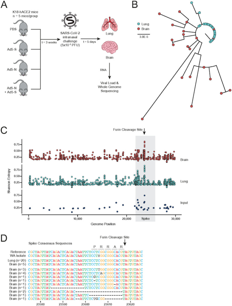
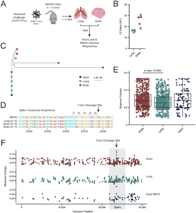
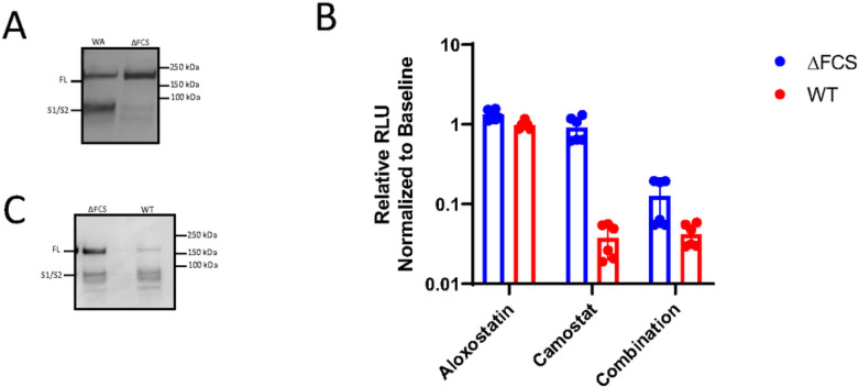
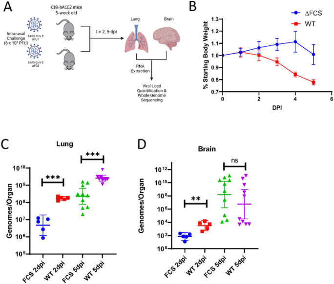
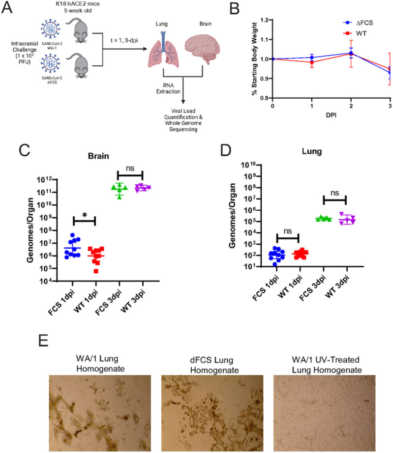
Severe COVID-19 and post-acute sequelae of SARS-CoV-2 infection are associated with neurological complications that may be linked to direct infection of the central nervous system (CNS), but the selective pressures ruling neuroinvasion are poorly defined. Here, we assessed SARS-CoV-2 evolution in the lung versus CNS of infected mice. Higher levels of viral diversity were observed in the CNS than the lung after intranasal challenge with a high frequency of mutations in the Spike furin cleavage site (FCS). Deletion of the FCS significantly attenuated virulence after intranasal challenge, with lower viral titers and decreased morbidity compared to the wild-type virus. Intracranial inoculation of the FCS-deleted virus, however, was sufficient to restore virulence. After intracranial inoculation, both viruses established infection in the lung, but this required reversion of the FCS deletion. Cumulatively, these data suggest a critical role for the FCS in determining SARS-CoV-2 tropism and compartmentalization with possible implications for the treatment of neuroinvasive COVID-19.

Keywords: COVID-19; Central Nervous System (CNS); Furin Cleavage Site (FCS); NeuroCOVID; SARS-CoV-2; Viral Evolution.


Conflict of interest statement

COMPETING FINANCIAL INTERESTS J.F.H. has received research support, paid to Northwestern University, from Gilead Sciences and is a paid consultant for Merck. All other authors declare no conflicts of interest.

Figures

Figure 1
Figure 1. Viral RNA from the CNS has a higher degree of genetic diversity regardless of vaccination status.
a, K18-hACE2 mice were vaccinated with Ad5 vaccines encoding spike, nucleocapsid, spike + nucleocapsid, or PBS as a negative control (n = 5 per group) three weeks before intranasal challenge with SARS-CoV-2. Brain and lung tissue were harvested 5 dpi. b, Phylogenetic tree of the consensus SARS-CoV-2 whole genome sequences (red tips = brain isolate, light blue tips = lung isolate). Branch length reflects nucleotide substitutions per site. c, Shannon entropy per position across the genome for the viral inoculate (n = 1, lower, dark blue), the lung isolates (n = 20, middle, light blue), and the brain isolates (n = 20, upper, red). The coding region for the spike protein is back shaded in gray with the FCS position labelled. d, Alignment of each consensus sequence across the Spike FCS region.
Figure 2
Figure 2. BALB/c mice infected with mouse-adapted SARS-CoV-2 have increased viral diversity in the CNS.
a, BALB/c mice were intranasally challenged with mouse-adapted SARS-CoV-2 (n = 5 mice). Brain and lung tissue were harvested 5 dpi. (n=5). b, Ct values of the SARS-CoV-2 viral RNA (N1 target) isolated from the brain and lung of each mouse as quantified by RT-qPCR. The black line indicates the average Ct value per compartment. c,Phylogenetic tree of the consensus SARS-CoV-2 whole genome sequences (dark blue tip = inoculate, red tips = brain isolate, light blue tips = lung isolate). Branch length reflects nucleotide substitutions per site. d, Alignment of each consensus sequence across the Spike FCS region. e, Box plots of the Shannon entropy at each position across the genome for the brain isolates (n = 5, red), the lung isolates (n = 5, light blue), and the viral inoculate (n = 1, dark blue). Box plots represent the median and interquartile range. A log-transformed linear mixed effects model was used to test for significant differences in overall genetic entropy between tissues. f, Shannon entropy per position across the genome for the viral inoculate (n = 1, lower, dark blue), the lung isolates (n = 5, middle, light blue), and the brain isolates (n = 5, upper, red).
Figure 3
Figure 3. Mutation of the FCS blocks Spike S1/S2 cleavage and mitigates utilization of an alternative entry pathway.
a, Pseudoviruses encoding for luciferase were generated with the WA-1 or ΔFCS deletion version of spike. Protein was isolated from pseudovirus cultures and subject to immunoblot staining against the spike protein. Shown is a representative blot of three independent western blots. b, Vero-E6 cells expressing hACE2 and TMPRSS2 were infected with WT or ΔFCS pseudotyped virus in the presence of presence of no drug, aloxostatin, camostat mesylate, or both. After 48 hours of infection, cells were lysed, and luciferase expression was quantified. Relative luciferase expression was normalized to untreated cells. Each condition was repeated three times. c, WT and ΔFCS virus were derived from infectious clones. Protein was isolated from viral stocks and subject to immunoblot staining against the spike protein. Shown is a representative blot of three independent western blots.
Figure 4
Figure 4. ΔFCS virus is attenuated after intranasal infection.
a, K18-hACE2 mice were intranasally inoculated with WA-1 and ΔFCS stocks. Mice were intranasally challenged with 6×10³ PFU of wild-type (red/purple) or ΔFCS (blue/green) SARS-CoV-2 and b, evaluated for weight loss. (n = 15 for each virus). Viral genomes/organ are plotted from the c, lung and d, brain was isolated and quantified via RT-qPCR. At 2 days post-infection (dpi), n = 5 mice per virus were euthanized with remaining n = 10 euthanized at 5 dpi. Statistical significance is denoted by asterisks, as compared to WT virus (**p < 0.005; ***p < 0.0005; Unpaired T test).
Figure 5
Figure 5. ΔFCS virus replicates faster in the CNS after intracranial inoculation.
a, K18-hACE2 mice were intracranially challenged with 102 PFU of wild-type (red/purple) or ΔFCS (blue/green) SARS-CoV-2. b, Mice were evaluated for weight loss (n = 15 for each virus). At 1 day post-infection (dpi), n = 10 mice per virus were euthanized with remaining n = 5 euthanized at 3 dpi. Viral RNA from the c, brain and d, lung was isolated and quantified via RT-qPCR. Viral genomes/organ are plotted. Statistical significance is denoted by asterisks, as compared to WT virus (*p < 0.05 Unpaired T test). e, Lung homogenate at 3dpi from WT or dFCS infected mice were inoculated onto a monolayer of Vero-E6 cells and incubated for three days. As a control, lung homogenate was UV treated to inactivate infectious virus in one group. Brightfield images of the monolayer is shown.
Figure 6
Figure 6. There is selective pressure for the deletion of the FCS in the CNS.
a, Phylogenetic tree of the consensus SARS-CoV-2 whole genome sequences after intranasal or intracranial challenge with WT or ΔFCS virus (dark blue tip = inoculate, red tips = intracranial challenge, green tips = intranasal challenge, triangle = brain isolate, square = lung isolate). Branch length reflects nucleotide substitutions per site. b, Alignment of each consensus sequence across the Spike FCS region after challenge with the WT (left) or ΔFCS (right) virus. c,Phylogenetic tree of SARS-CoV-2 quasispecies after intranasal or intracranial challenge with WT (left) or ΔFCS virus (right) (dark blue tip = inoculate, red tips = intracranial challenge, green tips = intranasal challenge, triangle = brain isolate, square = lung isolate). Branch length reflects nucleotide substitutions per site and tip size reflects the frequency of a given subpopulation in an isolate.

References

    1. Chi W.-Y. et al. COVID-19 vaccine update: vaccine effectiveness, SARS-CoV-2 variants, boosters, adverse effects, and immune correlates of protection. J. Biomed. Sci. 29, 82 (2022). - PMC - PubMed
    1. Hu B., Guo H., Zhou P. & Shi Z.-L. Characteristics of SARS-CoV-2 and COVID-19. Nat. Rev. Microbiol. 19, 141–154 (2021). - PMC - PubMed
    1. Stein S. R. et al. SARS-CoV-2 infection and persistence in the human body and brain at autopsy. Nature 612, 758–763 (2022). - PMC - PubMed
    1. Hartard C. et al. Multiorgan and Vascular Tropism of SARS-CoV-2. Viruses 14, (2022). - PMC - PubMed
    1. Gupta A. et al. Extrapulmonary manifestations of COVID-19. Nat. Med. 26, 1017–1032 (2020). - PMC - PubMed

Publication types

LinkOut - more resources