[Preprint]. 2023 Sep 23:2023.09.22.558020.
doi: 10.1101/2023.09.22.558020.

Role Of The C-C Motif Chemokine Ligand 5 (CCL5) And Its Receptor, C-C Motif Chemokine Receptor 5 (CCR5) In The Genesis Of Aldosterone-induced Hypertension, Vascular Dysfunction, And End-organ Damage

Affiliations

Role Of The C-C Motif Chemokine Ligand 5 (CCL5) And Its Receptor, C-C Motif Chemokine Receptor 5 (CCR5) In The Genesis Of Aldosterone-induced Hypertension, Vascular Dysfunction, And End-organ Damage

Rafael M Costa et al. bioRxiv. .

Update in

Abstract

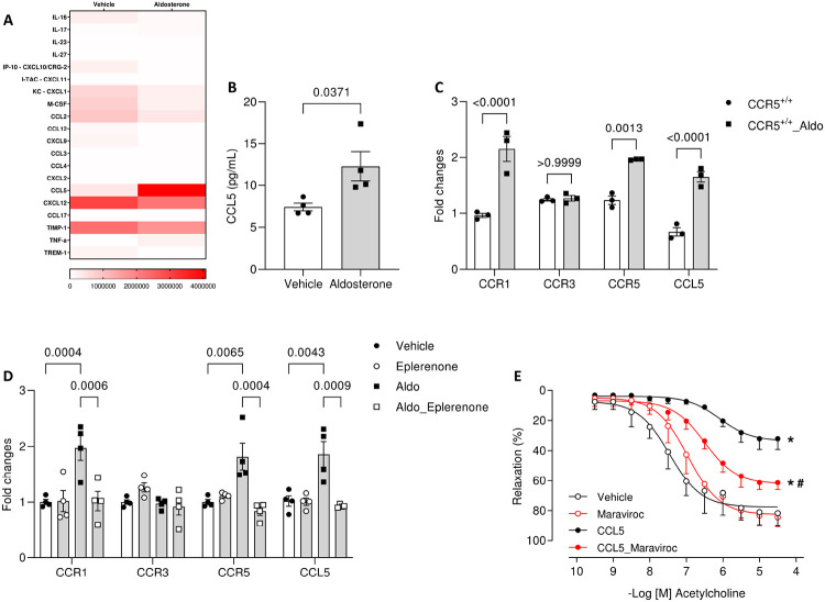
Background: Aldosterone, a mineralocorticoid steroid hormone, has been described to initiate cardiovascular diseases by triggering exacerbated sterile vascular inflammation. The functions of C-C Motif Chemokine Ligand 5 (CCL5) and its receptor, C-C Motif Chemokine Receptor 5 (CCR5), are well known in infectious diseases, but their roles in the genesis of aldosterone-induced vascular injury and hypertension are unknown.

Methods: We analyzed the vascular profile, blood pressure, and renal damage in wild-type (CCR5+/+) and CCR5 knockout (CCR5-/-) mice treated with aldosterone (600 μg/kg/day for 14 days) while receiving 1% saline to drink.

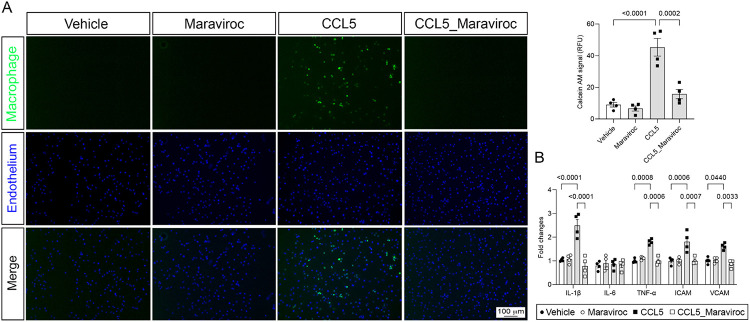
Results: Here, we show that CCR5 plays a central role in aldosterone-induced vascular injury, hypertension, and renal damage. Long-term infusion of aldosterone in CCR5+/+ mice resulted in exaggerated CCL5 circulating levels and vascular CCR5 expression. Aldosterone treatment also triggered vascular injury, characterized by endothelial dysfunction and inflammation, hypertension, and renal damage. Mice lacking CCR5 were protected from aldosterone-induced vascular damage, hypertension, and renal injury. Mechanistically, we demonstrated that CCL5 increased NADPH oxidase 1 (Nox1) expression, reactive oxygen species (ROS) formation, NFκB activation, and inflammation and reduced nitric oxide production in isolated endothelial cells. These effects were abolished by antagonizing CCR5 with Maraviroc. Finally, aortae incubated with CCL5 displayed severe endothelial dysfunction, which is prevented by blocking Nox1, NFκB, or with Maraviroc treatment.

Conclusions: Our data demonstrate that CCL5/CCR5, through activation of NFkB and Nox1, is critically involved in aldosterone-induced vascular and renal damage and hypertension. Our data place CCL5 and CCR5 as potential targets for therapeutic interventions in conditions with aldosterone excess.

Keywords: NADPH oxidases; aldosterone; chemokines; chemokines receptors; oxidative stress.


Figures

Figure 1.
Figure 1.. Aldosterone-induced CCL5 high levels promotes endothelial dysfunction.
Inflammatory profile in plasma, measured by proteome profiler mouse cytokine array and presented as heat map (A); CCL5 levels, measured by ELISA (B); and chemokine receptors expression, measured by RT-PCR (C), from CCR5+/+ mice treated with vehicle or aldosterone (600 μg/kg/day for 14 days). Chemokine receptors expression, measured by RT-PCR, in endothelial cells (MEC) treated with vehicle or aldosterone (0.1μM) in the presence of Eplerenone (1μM) (D). Concentration-effect curves to acetylcholine in aortae from CCR5+/+ mice incubated with vehicle or CCL5 (100ng/ml, 24h) in the presence of Maraviroc (40μM) (E). Values represent means ± SEM (n= 3-7). Student t test or ANOVA test. *p<0.05 vs. Vehicle; #p<0.05 vs. CCL5.
Figure 2.
Figure 2.. CCR5 deficiency prevents endothelial dysfunction and vascular inflammation induced by aldosterone.
Concentration-effect curves to acetylcholine (A) and sodium nitroprusside (B); phosphorylated (p65 subunit) and total NFκB expression, analyzed by western blot to (C); inflammatory markers, measured by RT-PCR (D), in aortae from CCR5+/+ and CCR5−/− mice treated with vehicle or aldosterone (600 μg/kg/day + saline for 14 days). Phosphorylated (p65 subunit) and total NFκB expression analyzed by western blot in aortae from CCR5+/+ mice incubated with vehicle or CCL5 (100ng/ml, 24h) (E) and concentration-effect curves to acetylcholine (F) in aortae from CCR5+/+ mice incubated with vehicle or CCL5 (100ng/ml, 24h) in the presence of BMS-345541 (5μM). Values represent means ± SEM (n= 4-7). Student t test or ANOVA test. *p<0.05 vs. CCR5+/+ or Vehicle; #p<0.05 vs. CCR5+/+_Aldo or CCL5.
Figure 3.
Figure 3.. CCL5 via CCR5 induces endothelial cell activation and immune cell adhesion.
Photomicrography and fluorescence intensity depicting labeled macrophages (calcein-AM probe, green) and endothelial cells (MEC, DAPI, blue). MEC were treated with vehicle, Maraviroc (40μM, 30 minutes prior CCL5 incubation), CCL5 (100ng/ml, 24h) or CCL5 plus Maraviroc-stimulated MEC. Scale bar = 100μm (A). Inflammatory markers, measured by RT-PCR, in MEC treated with vehicle or CCL5 (100ng/ml) in the presence of Maraviroc (40μM) (B). Values represent means ± SEM (n= 4). ANOVA test.
Figure 4.
Figure 4.. Aldosterone via CCR5 promotes increased blood pressure and kidney injury.
Systolic blood pressure (A), diastolic blood pressure (B), and mean arterial pressure (C), measured via radiotelemetry, in CCR5+/+ and CCR5−/− mice treated with vehicle or aldosterone (600 μg/kg/day and saline for 14 days). Immunofluorescence for podocyte marker synaptopodin (Synap; green), endothelial marker endomucin (Endom; red), and nuclei (DAPI, blue) represented by letters A to D, α-smooth muscle actin (α-SMA; green) and nuclei (DAPI, blue) represented by letters E to H, fibrosis marker and proximal tubules were visualized with fluorescein-labeled collagen III (red) and Lotus tetragonolobus lectin (LTA; green), which are represented by letters I to L. Images were obtained from kidney sections of CCR5+/+ and CCR5−/− mice treated with vehicle or aldosterone. The images shown are representative of three independent experiments. Scale bar = 20 or 50μm (D). Kidney injury markers, measured by RT-PCR in CCR5+/+ and CCR5−/− mice treated with vehicle or aldosterone (E), and proteinuria levels in CCR5+/+ and CCR5−/− mice treated with vehicle or aldosterone measured by 10% SDS-PAGE gels followed by staining with Coomassie Brilliant. Albumin was used as a control analyzed. Values represent means ± SEM (n= 3-7). Student t test or ANOVA test. *p<0.05 vs. CCR5+/+.
Figure 5.
Figure 5.. Aldosterone, via CCR5, promotes endothelial dysfunction by NOX1-dependent mechanisms.
Representative western blot (A) to NOX1 (i), NOX2 (ii) and NOX4 (iii) expression; ROS generation, measured Amplex red assay (B); and concentration-effect curves to acetylcholine, in the presence of NOXA1ds (10μM) (C), in aortae from CCR5+/+ and CCR5−/− mice treated with vehicle or aldosterone (600 μg/kg/day for 14 days). Representative western blot to NOX1 (i); ROS generation, measured by Amplex red assay (ii) (D); and concentration-effect curves to acetylcholine, in the presence of NOXA1ds (10μM) (E), in aortae from CCR5+/+ mice incubated with vehicle or CCL5 (100ng/ml). Representative western blot to NOX1 (i) and ROS generation, measured by Amplex red (ii), in endothelial cells (MEC) treated with vehicle or CCL5 (100ng/ml) in the presence of Maraviroc (40μM) and NOXA1ds (10μM) (F). Values represent means ± SEM (n= 4-7). ANOVA test. *p<0.05 vs. CCR5+/+ or Vehicle; #p<0.05 vs. CCR5+/+_Aldo or CCL5.
Figure 6.
Figure 6.. CCL5 induces an increase in NOX1 expression and NFKB activity by a positive feedback mechanism in endothelial cells.
Representative western blot to NOX1 expression (A) and ROS generation, measured by Amplex red (B), in endothelial cells (MEC) treated with vehicle or CCL5 (100ng/ml), in the presence of BMS-345541 (5μM). Representative western blot to phosphorylated and total NFκB expression in MEC treated with vehicle or CCL5, in the presence of NOXA1ds (10μM) (C). Values represent means ± SEM (n= 4). ANOVA test.

References

    1. Scott JH, Menouar MA, Dunn RJ. Physiology, aldosterone. Statpearls. Treasure Island (FL); 2023. - PubMed
    1. Briones AM, Nguyen Dinh Cat A, Callera GE, Yogi A, Burger D, He Y, Correa JW, Gagnon AM, Gomez-Sanchez CE, Gomez-Sanchez EP, Sorisky A, Ooi TC, Ruzicka M, Burns KD, Touyz RM. Adipocytes produce aldosterone through calcineurin-dependent signaling pathways: Implications in diabetes mellitus-associated obesity and vascular dysfunction. Hypertension. 2012;59:1069–1078 - PubMed
    1. Dinh Cat AN, Friederich-Persson M, White A, Touyz RM. Adipocytes, aldosterone and obesity-related hypertension. J Mol Endocrinol. 2016;57:F7–F21 - PubMed
    1. Calhoun DA. Aldosterone and cardiovascular disease: Smoke and fire. Circulation. 2006;114:2572–2574 - PubMed
    1. He BJ, Anderson ME. Aldosterone and cardiovascular disease: The heart of the matter. Trends Endocrinol Metab. 2013;24:21–30 - PMC - PubMed

Publication types