[Preprint]. 2023 Sep 22:2023.09.22.558671.
doi: 10.1101/2023.09.22.558671.

Cryo-EM structure of Alzheimer's disease tau filaments with PET ligand MK-6240

Affiliations

Cryo-EM structure of Alzheimer's disease tau filaments with PET ligand MK-6240

Peter Kunach et al. bioRxiv. .

Update in

  • Cryo-EM structure of Alzheimer's disease tau filaments with PET ligand MK-6240.
    Kunach P, Vaquer-Alicea J, Smith MS, Monistrol J, Hopewell R, Moquin L, Therriault J, Tissot C, Rahmouni N, Massarweh G, Soucy JP, Guiot MC, Shoichet BK, Rosa-Neto P, Diamond MI, Shahmoradian SH. Kunach P, et al. Nat Commun. 2024 Oct 1;15(1):8497. doi: 10.1038/s41467-024-52265-x. Nat Commun. 2024. PMID: 39353896 Free PMC article.

Abstract

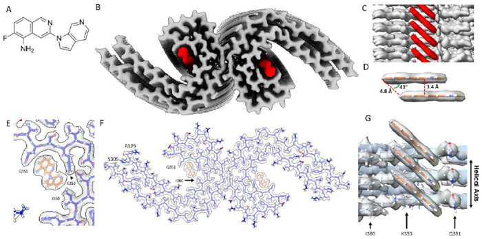
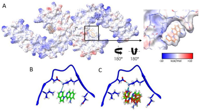
Positron Emission Tomography (PET) ligands have advanced Alzheimer's disease (AD) diagnosis and treatment. Using autoradiography and cryo-EM, we identified AD brain tissue with elevated tau burden, purified filaments, and determined the structure of second-generation high avidity PET ligand MK-6240 at 2.31 Å resolution, which bound at a 1:1 ratio within the cleft of tau paired-helical filament (PHF), engaging with glutamine 351, lysine K353, and isoleucine 360. This information elucidates the basis of MK-6240 PET in quantifying PHF deposits in AD and may facilitate the structure-based design of superior ligands against tau amyloids.

Keywords: Alzheimer’s disease (AD); Cryo-electron microscopy (cryo-EM); Neurofibrillary tangles (NFTs); Positron emission tomography (PET); Tau; [18F]MK-6240 PET tracer; atomic resolution; autoradiography; binding affinity; binding agents; in vivo PET imaging; pi-pi aromatic stacking.


Conflict of interest statement

Competing Interests The authors declare no competing interests.

Figures

Figure. 1:
Figure. 1:. Structure of MK-6240 bound to tau PHF.
(A) Molecular structure of MK-6240. (B) Cryo-EM map of AD tau PHFs (Grey) with bound MK-6240 (Red). (C) Side-view depiction of MK-6240 (red) situated within the binding pocket of the cleft within the AD tau PHF (Grey). MK-6240 adopted a stacked arrangement perpendicular to the fibril axis. One MK-6240 molecule spanned approximately two tau monomer rungs. (D) Isolated atomic representation of MK-6240 with its binding orientation. Peripheral atoms (alpha-carbon Hydrogen, left; and Fluorine, right) exhibit a 4.8 Å distance between them. Between heterocycles or parallel atoms within MK-6240 molecules, a ~3.3 Å distance is observed. (E) Close-up depiction of the binding site, accentuating the proximity of the three amino acids (Q351, K353, and I360) to MK-6240. (F) Atomic model showcasing the MK-6240 binding site on paired helical filaments. Cryo-EM density (white) is juxtaposed with the atomic model of the tau fold (blue) and MK-6240 (orange). (G) Side-view, zoomed- in perspective (from within the cleft) of the MK-6240 binding site, revealing the ~1:1 stoichiometry and the angle that aligns individual MK-6240 molecules with both themselves and the filament axis.
Figure. 2:
Figure. 2:. Electrostatic interactions between MK-6240 and PHFs and correspondence of the docking prediction to the experimental result.
(A) Coulombic energy map depicting the cryo-EM density of the binding pocket, with amino acids from the tau fold and MK-6240. (B) Optimal pose resulting from MK-6240 binding via symmetry docking, revealing hydrogen bonding involving the secondary amine of the azaindole ring and K353. Minimized energetic scores align with the manually docked pose of MK-6240 in the cryo-EM density as shown in (B). (C) The top four poses (Green > Yellow > Orange > Red) generated through symmetry docking, showcasing robust alignment and a predilection for the orientation of the secondary amine of the azaindole ring system to facilitate interaction with K353. Flexibility variations toward the solvent-exposed region are observed among these poses.

References

    1. Jack C. R. et al. A/T/N: An unbiased descriptive classification scheme for Alzheimer disease biomarkers. Neurology 87, 539–547 (2016). - PMC - PubMed
    1. Jack C. R. Jr. et al. NIA-AA Research Framework: Toward a biological definition of Alzheimer’s disease. Alzheimer’s & Dementia 14, 535–562 (2018). - PMC - PubMed
    1. Hyman B. T. et al. National Institute on Aging-Alzheimer’s Association guidelines for the neuropathologic assessment of Alzheimer’s disease. Alzheimers Dement 8, 1–13 (2012). - PMC - PubMed
    1. Yagishita S., Itoh Y., Nan W. & Amano N. Reappraisal of the fine structure of Alzheimer’s neurofibrillary tangles. Acta Neuropathologica 54, 239–246 (1981). - PubMed
    1. Crowther R. A. Straight and paired helical filaments in Alzheimer disease have a common structural unit. Proceedings of the National Academy of Sciences 88, 2288–2292 (1991). - PMC - PubMed

Publication types