[Preprint]. 2023 Sep 18:rs.3.rs-3269310.
doi: 10.21203/rs.3.rs-3269310/v1.

Sde Proteins Coordinate Ubiquitin Utilization and Phosphoribosylation to Promote Establishment and Maintenance of the Legionella Replication Vacuole

Affiliations

Sde Proteins Coordinate Ubiquitin Utilization and Phosphoribosylation to Promote Establishment and Maintenance of the Legionella Replication Vacuole

Ralph Isberg et al. Res Sq. .

Update in

Abstract

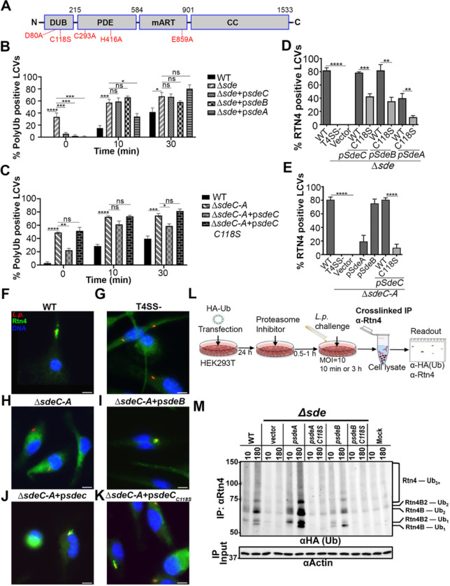
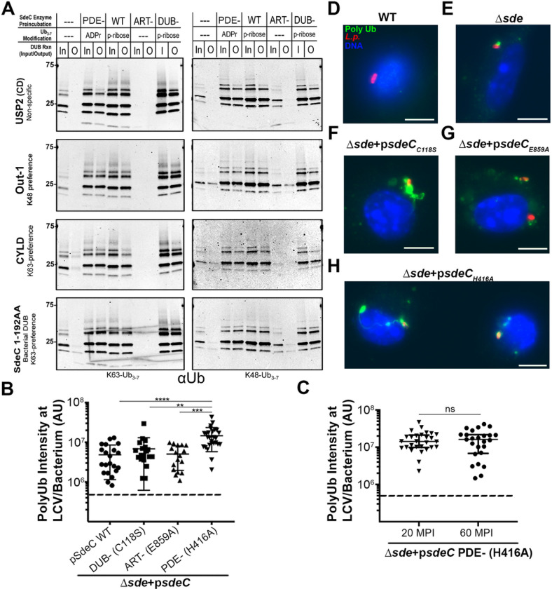
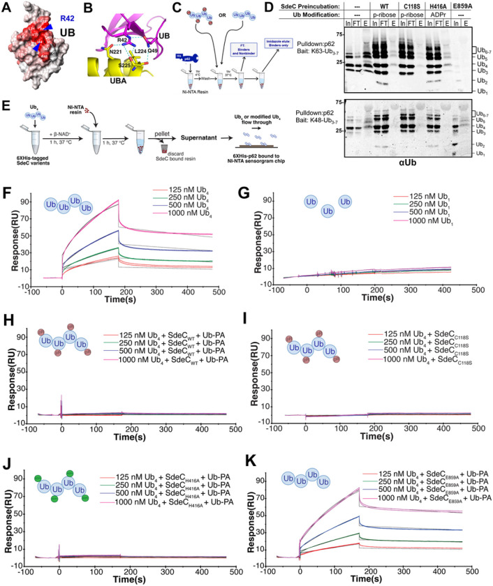
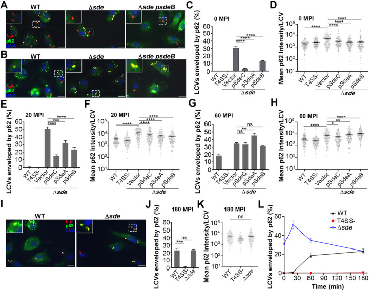
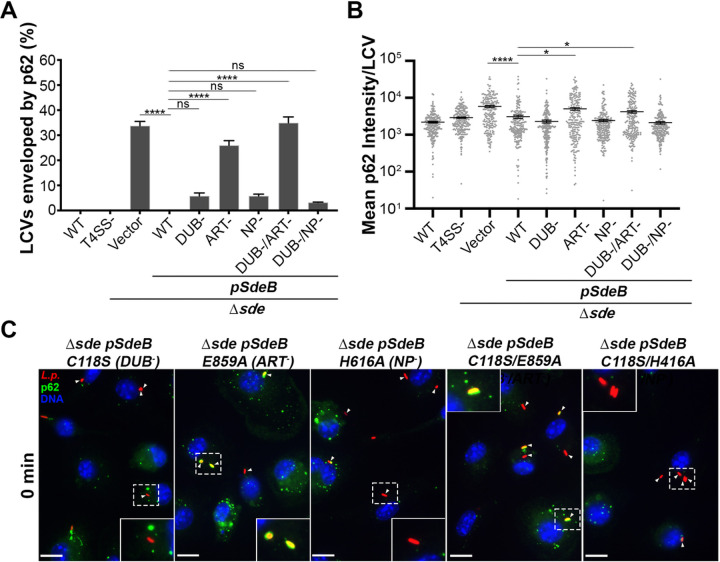
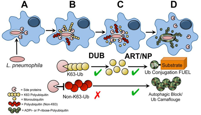
The Legionella pneumophilaSde family of translocated proteins promote host tubular endoplasmic reticulum (ER) rearrangements that are tightly linked to phosphoribosyl-ubiquitin (pR-Ub) modification of Reticulon 4 (Rtn4). Sde proteins have two additional activities of unclear relevance to the infection process: K63 linkage-specific deubiquitination and phosphoribosyl modification of polyubiquitin (pR-Ub). We show here that the deubiquitination activity (DUB) stimulates ER rearrangements while pR-Ub protects the replication vacuole from cytosolic surveillance by autophagy. Loss of DUB activity was tightly linked to lowered pR-Ub modification of Rtn4, consistent with the DUB activity fueling the production of pR-Ub-Rtn4. In parallel, phosphoribosyl modification of polyUb, in a region of the protein known as the isoleucine patch, caused an absolute block in binding by the autophagy adapter p62. An inability of Sde mutants to modify polyUb resulted in immediate p62 association, a critical precursor to autophagic attack. The ability of Sde WT to block p62 association decayed quickly after bacterial infection, as predicted by the presence of previously characterized L. pneumophila effectors that inactivate Sde and remove polyUb. In sum, these results show that the accessory Sde activities act to stimulate ER rearrangements and protect from host innate immune sensing in a temporal fashion.

Keywords: Legionella; autophagy; deubiquitination; intracellular replication; macrophages; ubiquitination.


Figures

Figure 1
Figure 1. The Sde DUB domain is required for efficient phosphoribose-ubiquitination of Rtn4.
(A) Domain structure of Sde family proteins. Catalytically inactive point mutations are shown in red. (B, C) Percent poly-Ub positive LCVs of L. pneumophila strains at noted timepoints post-infection. (D, E)Quantification of Rtn4-positive LCVs. BMDMs were challenged with indicated strains for the noted infection times, followed by fixation, permeabilization with 1% Triton X-100 and probing as described in Materials and Methods. 0 MPI was immediately after 5 min centrifugation of bacteria onto BMDM. Noted strains are wild type, Lp02; Dsde, DsidEDsdeCDsdeB-A; DsdeC-A, Dlpg2153–2157 (KK034), listed in Supplemental Table 1. At least 150 LCVs per experiment were evaluated and data were pooled from 3 biological replicates (mean ± SEM; one-way ANOVA with Dunnett’s multiple comparison (B, C)and Student’s t-test (D, E); ns (non-significant), *p< 0.0001). (F-K) A representative micrograph of Rtn4 recruitment, scale bar 5 μm. Macrophages were challenged with either WT or noted mutant strains for 1 hr, fixed, permeabilized and probed with anti-L. pneumophila (Alexa Fluor 594 secondary, red), anti-Rtn4 (Alexa Fluor 488 secondary, green), and Hoechst (nucleus, blue). (L) Cartoon of strategy to identify pR-Ub modified Rtn4. (M) Immunoblot image of anti-Rtn4 immunoprecipitates by immunoprobing with anti-HA. To right of immunoblots are Rtn4 isoforms and modification status. HEK293T cells were challenged with noted L. pneumophila strains for either 10 or 180 mins. Modified Rtn4 substrates were immunoprecipitated using crosslinked anti-Rtn4, fractionated on 7.5% SDS-PAGE, and probed with anti-HA. Lanes: WT, Lp02; vector, Dsde; psdeA, Dsde expressing SdeA; psdeA C118S, Dsde expressing SdeA DUB mutant; psdeB, Dsde expressing SdeB; psdeB C118S, Dsde expressing SdeB DUB mutant; MOCK, uninfected.
Figure 2
Figure 2. The ART domain blocks DUB activity and promotes hyper-polyubiquitination of the LCV.
(A). SdeC WT or its catalytic mutant derivatives (1μg) were adsorbed to Ni-NTA resin for 1 hr at 4°C, then incubated with either K63 or K48-linked polyUb3–7 (2μg) for 2 hrs at 37°C. SdeC resin was removed and polyUb3–7 was then incubated with: human recombinant Polyhistidine-otubain 1, isoform 1 (100 nM); recombinant USP2 catalytic domain (50 nM); recombinant Polyhistidine-CYLD (50 nM); or recombinant SdeC DUB CD 1–192 (50 nM) at 37°C for 2 hrs. Cleavage was monitored by immunoblotting with anti-Ub (FK1). (B) Quantification of polyUb intensity associated with individual LCVs of the noted strains at 20 MPI. (C) PolyUb intensity of LCVs harboring Dsde expressing SdeC PDE (H416A) mutant at 20 or 60 MPI. Data are shown in median ± 25%ile; Mann-Whitney test; ns (non-significant), **p<0.01, ***p<0.001, ****p < 0.0001. The dotted lines represent background level of polyUb intensity about LCVs. (D-H) Examples of polyUb-positive LCVs. BMDMs were challenged by 5 min centrifugation at an MOI of 1, incubated with noted L. pneumophila strains, fixed, permeabilized with 1% Triton X100, and probed with anti-polyUb (Alexa Fluor 488 secondary, green), anti-L. pneumophila (Alexa Fluor 594 secondary, red), and Hoechst (blue). Strains used: WT, Lp02; Δsde, ΔsidE ΔsdeC ΔsdeB-A; Δsde psdeCWT, Dsde expressing SdeC WT; Δsde psdeCC118S, Dsde expressing SdeC DUB mutant; Δsde psdeCE859A, Dsde expressing SdeC ART mutant; Δsde psdeCH416A, Dsde expressing SdeC PDE mutant. Scale bar: 5mm.
Figure 3
Figure 3. Sde-mediated modification of polyUb prevents its recognition by p62.
(A) Space filling model of Ub. R42 residue (blue letter) and I44 residue are indicated with blue arrows (from reference, ).(B) Interface residues between UBA domain and Ub, showing possible involvement of Ub R42 in interacting with UBA domain (from reference, ). (C, D) Ub modification at R42 blocks p62 binding (Materials and Methods). Ni-NTA-p62 was incubated with Ub3–7 that had been treated with SdeC WT or its catalytic mutant derivatives and allowed to bind (In). Unbound Ub3–7 was removed (FT), the beads were washed, and then eluted with 250mM imidazole (E: elution). The p62-bound poly-Ub in the eluate was fractionated by SDS-PAGE followed by probing with anti-Ub. Relative loading: In, input (2.5%); FT, flow-through (2.5%); E, eluate (25%). (E) Surface plasmon resonance (SPR) quantitation of p62 binding to Ub derivatives. Ub1 or Ub4 was incubated with SdeC WT or its derivatives immobilized on Ni-NTA resin, the supernatant containing modified Ub derivatives was incubated with 6xHis-tagged p62 bound to a Ni-NTA chip, and binding affinity was measured by plasmon resonance. (F, G) Sensorgrams of Ub4 or Ub1 binding to p62. Binding of Ub4 to p62 yields a KD= 0.97 × 10⁻⁷ M. (H-K) Sensorgrams of Ub4, modified as noted in panels (red balls: phosphoribosyl addition; green balls: ADP-ribosyl addition). Reactions were formed in the presence of Ub-propargylamide (DUB inhibitor).
Figure 4
Figure 4. Transient blockade of p62 recruitment to the LCV in the presence of Sde family members.
(A-B) Examples of punctate (A) or enveloped (B) p62 morphology localized around LCVs after challenge of BMDMs with noted L. pneumophila strains. The scale bar represents 10 μm. (C, E, G, J) The Percentage of circumferential p62 associated with LCVs at 0 (immediate bacterial contact; C) or 20 (E) or 60 (G) or 180 (J) MPI. 100 LCVs per replicate were counted in 3 biological replicates (mean ± SEM; one-way ANOVA with Dunnett’s multiple comparison; ns (non-significant), **p<0.01, ***p<0.001, ****p < 0.0001). (D, F, H, K) Quantification of mean p62 intensity/pixel around LCVs at 0 (D) or 20 (F) or 60 (H) or 180 (K) MPI. At least 70 vacuoles per replicate were quantified and data were pooled from 3 biological replicates (mean ± SEM; one-way ANOVA with Dunnett’s multiple comparison (D, F, H) and Student’s t-test (K); ns (non-significant), *p<0.05, **p<0.01, ****p < 0.0001). (I) A representative micrograph of p62 association with LCVs at 180 MPI, scale bar 10 μm. (L) Kinetics of blockade against p62 recruitment. Data acquired from panels C, E, G and J, and shown as mean ± SEM.
Figure 5
Figure 5. ART activity is sufficient to block p62 recruitment to the replication vacuole.
(A) Quantification of individual LCVs enveloped by p62 at 0 MPI. BMDMs were challenged with the noted Legionella strains. (B) Quantitation of mean p62 intensity/pixel associated with individual LCVs. Images of individual vacuoles were grabbed and pixel intensities of p62 staining about regions of interest were determined (Materials and Methods). 210 LCVs were counted. Data are shown as means ± SEM (one-way ANOVA with Dunnett’s multiple comparison; ns (non-significant), *p<0.05, **p<0.01, ****p < 0.0001). (C)-Examples of recruited p62 to LCVs harboring Dsde expressing SdeC catalytic mutant derivatives. Scale bar: 10 mm.
Figure 6
Figure 6. Sde accessory activities drive Rtn4 rearrangements and block autophagic recognition.
The DUB activity provides fuel for pR-Ub modification of Rtn4, driving rapid ER-associated rearrangements. (A)Bacteria are internalized, Sde family proteins are translocated. (B) Polyubiquitination occurs about the replication vacuole. (C) Digestion of polyUb provides Ub fuel for ART/PDE domain to catalyze pR-Ub conjugation to substrates such as Rtn4. (D) Rtn4-pR-Ub modified protein accumulates about the replication vacuole polyUb is modified by phosphoribosylation. The pR-modification of the polyUb at R42 residue stabilizes polyubiquitination about LCVs, simultaneously preventing degradation by host cell-derived DUBs and blocking recognition by the autophagic machinery.

References

    1. Cunha B.A., Wu G. & Raza M. Clinical Diagnosis of Legionnaire’s Disease: Six Characteristic Clinical Predictors. Am J Med 128, e21–22 (2015). - PubMed
    1. Cunha B.A., Burillo A. & Bouza E. Legionnaires’ disease. Lancet 387, 376–385 (2016). - PubMed
    1. Safdar N., Crnich C.J. & Maki D.G. The pathogenesis of ventilator-associated pneumonia: its relevance to developing effective strategies for prevention. Respir Care 50, 725–739; discussion 739–741 (2005). - PubMed
    1. Horwitz M.A. & Silverstein S.C. Interaction of the Legionnaires’ disease bacterium (Legionella pneumophila) with human phagocytes. I. L. pneumophila resists killing by polymorphonuclear leukocytes, antibody, and complement. J Exp Med 153, 386–397 (1981). - PMC - PubMed
    1. Rowbotham T.J. Isolation of Legionella pneumophila from clinical specimens via amoebae, and the interaction of those and other isolates with amoebae. J Clin Pathol 36, 978–986 (1983). - PMC - PubMed

Publication types