Case Reports
. 2023 Sep 14:10:1251874.
doi: 10.3389/fcvm.2023.1251874. eCollection 2023.

Ongoing substrate-driven atrial fibrillation "boxed" in the left atrial posterior wall with ablation: a case report

Affiliations
Case Reports

Ongoing substrate-driven atrial fibrillation "boxed" in the left atrial posterior wall with ablation: a case report

Vassil Traykov et al. Front Cardiovasc Med. .

Abstract

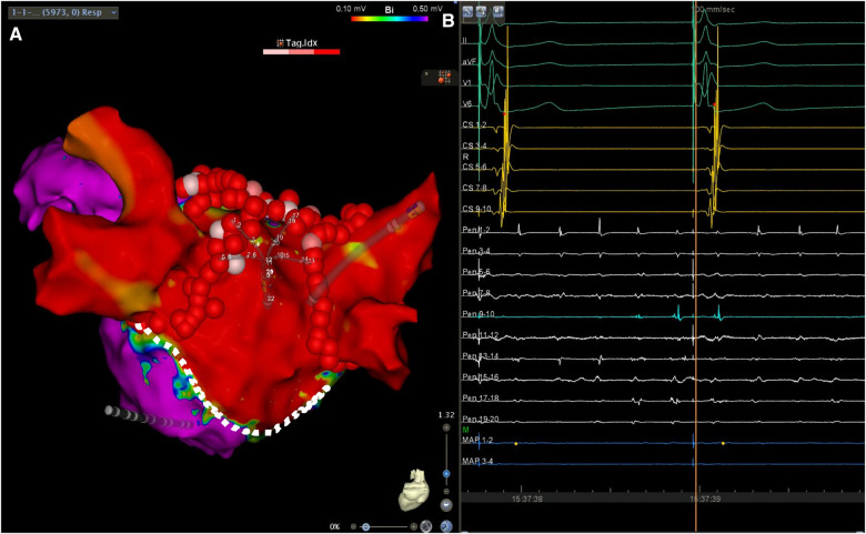
Patients undergoing valve surgery for rheumatic heart disease are expected to develop significant atrial arrhythmogenic substrates outside of the pulmonary veins, which sometimes require complex ablation techniques for the treatment of symptomatic arrhythmias. We describe, herein, the case of a 76-year-old male undergoing endocardial ablation for the treatment of symptomatic persistent atrial fibrillation which developed after aortic and mitral valve replacement with a simultaneous tricuspid ring annuloplasty. Following pulmonary vein isolation, the patient's atrial fibrillation was converted into cavotricuspid isthmus-dependent atrial flutter. After a successful cavotricuspid isthmus ablation, the arrhythmia reverted back to a left atrial tachyarrhythmia originating from the posterior wall. A linear left atrial lesion led to the electrical isolation of a large area, which included the posterior wall, as well as the containment of the ongoing fibrillatory activity, while sinus rhythm was restored in the rest of the atria. In conclusion, successful left atrial posterior wall isolation can be achieved in the setting of severe scarring due to previous atriotomy by creating a linear lesion on the atrial roof, in conjunction with pulmonary vein isolation, sparing the patient from requiring bottom-line ablation, and avoiding possible esophageal injury. Such compartmentalization of the left atrium may effectively contain local fibrillatory activity, while allowing for the restoration of sinus rhythm.

Keywords: ablation; box lesion; case report; independent tachycardia; posterior wall.


Conflict of interest statement

The authors declare that the research was conducted in the absence of any commercial or financial relationships that could be construed as a potential conflict of interest.

Figures

Figure 1
Figure 1
Left atrial voltage map in posteroanterior (A) and anteroposterior (B) views, created during AF demonstrating a large area of low voltage occupying mainly, but not exclusively, the LAPW. Color bar settings 0.1–0.24 mV as suggested by Rodríguez-Mañero et al. (4). AF, atrial fibrillation; LAPW, left atrial posterior wall.
Figure 2
Figure 2
(A) Biatrial map during atrial flutter demonstrating the presence of a clockwise peritricuspid re-entry circuit with passively activated LA. (B) Surface ECG leads I, II, V1 and V6, along with intracardiac recordings from the multipolar mapping catheter (Pen) located at the LAPW, the ablation catheter (Abl) located at the cavotricuspid isthmus, and a decapolar catheter in the CS. Note the change in the activation in the activation sequence in the CS. Sweep speed was 100 mm/s. LA, left atrium; ECG, electrocardiogram; CS, coronary sinus; other abbreviations as in Figure 1.
Figure 3
Figure 3
(A) Left atrial voltage map in anteroposterior view demonstrating the lesion set including circumferential lesions encircling both PV antra as well as a linear lesion at the LA roof. The asterisk denotes the location at which RF application resulted in LAPW isolation and arrhythmia termination. (B) Surface ECG leads I, II, V1 and V6, along with intracardiac recordings from the multipolar mapping catheter located at the LAPW (Pen), the mapping/ablation catheter, and the decapolar catheter in the coronary sinus. Note the termination of arrhythmia in the rest of the atria as seen on the coronary sinus catheter bipoles and ongoing atrial fibrillation/atrial flutter at the LAPW. Sweep speed was 100 mm/s. MAP, mapping/ablation catheter; RF, radiofrequency; other abbreviations as in Figures 1, 2.
Figure 4
Figure 4
(A) Left atrial voltage map in posteroanterior view demonstrating a wide area of low voltage occupying the LAPW. The lesion set consisting of circumferential antral PV isolation and a roof line lesion connecting the upper veins is also shown. The silhouette of the multipolar mapping catheter is shown to be located on the LAPW. (B) Surface ECG leads I, II, aVF, V1 and V6, along with intracardiac recordings from the multipolar mapping catheter (Pen) located at the LAPW, the mapping/ablation catheter, and the decapolar catheter in the coronary sinus. Note the ongoing atrial fibrillation/atrial flutter at the LAPW, while the rest of the atrium is activated following retrograde ventriculoatrial conduction by the pacemaker effectively stimulating the conduction system. Sweep speed was 100 mm/s. Sweep speed was 100 mm/s. The dotted line shows the presumed atriotomy line, which most likely aided LAPW isolation using a roof line only. Color bar settings 0.1–0.5 mV. Abbreviations as in Figures 1, 2.

References

    1. Kaba RA, Momin A, Camm J. Persistent atrial fibrillation: the role of left atrial posterior wall isolation and ablation strategies. J Clin Med. (2021) 10:3129. 10.3390/jcm10143129 - DOI - PMC - PubMed
    1. Thiyagarajah A, Kadhim K, Lau DH, Emami M, Linz D, Khokhar K, et al. Feasibility, safety, and efficacy of posterior wall isolation during atrial fibrillation ablation: a systematic review and meta-analysis. Circ Arrhythm Electrophysiol. (2019) 12:e007005. 10.1161/CIRCEP.118.007005 - DOI - PubMed
    1. John B, Stiles MK, Kuklik P, Chandy ST, Young GD, Mackenzie L, et al. Electrical remodelling of the left and right atria due to rheumatic mitral stenosis. Eur Heart J. (2008) 29:2234–43. 10.1093/eurheartj/ehn329 - DOI - PubMed
    1. Rodríguez-Mañero M, Valderrábano M, Baluja A, Kreidieh O, Martínez-Sande J, García-Seara J, et al. Validating left atrial low voltage areas during atrial fibrillation and atrial flutter using multielectrode automated electroanatomic mapping. JACC Clin Electrophysiol. (2018) 4(12):1541–52. 10.1016/j.jacep.2018.08.015 - DOI - PubMed
    1. Wielandts JY, Kyriakopoulou M, Almorad A, Hilfiker G, Strisciuglio T, Phlips T, et al. Prospective randomized evaluation of high power during CLOSE-guided pulmonary vein isolation: the POWER-AF study. Circ Arrhythm Electrophysiol. (2021) 14:e009112. 10.1161/CIRCEP.120.009112 - DOI - PubMed

Publication types

LinkOut - more resources